3LJQ image
Deposition Date 2010-01-26
Release Date 2010-11-24
Last Version Date 2023-09-06
Entry Detail
PDB ID:
3LJQ
Keywords:
Title:
Crystal Structure of the Glycosylasparaginase T152C apo-precursor
Biological Source:
Source Organism:
Host Organism:
Method Details:
Experimental Method:
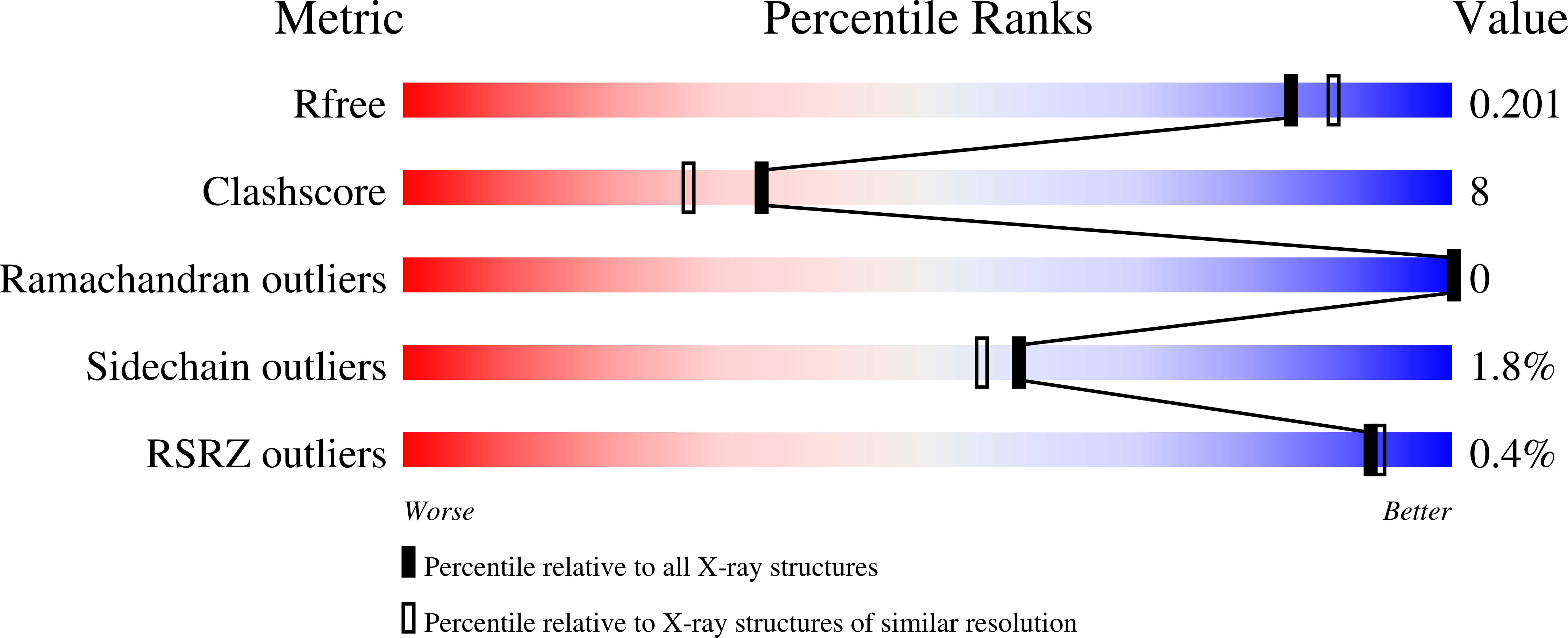
Resolution:
1.90 Å
R-Value Free:
0.19
R-Value Work:
0.15
R-Value Observed:
0.15
Space Group:
P 1
Macromolecular Entities
Polymer Type:polypeptide(L)
Molecule:N(4)-(Beta-N-acetylglucosaminyl)-L-asparaginase
Mutations:T152C
Chain IDs:A, B (auth: C)
Chain Length:299
Number of Molecules:2
Biological Source:Flavobacterium meningosepticum
Primary Citation
Crystallographic snapshot of glycosylasparaginase precursor poised for autoprocessing.
J.Mol.Biol. 403 120 130 (2010)
PMID: 20800597 DOI: 10.1016/j.jmb.2010.08.038

Abstact

Glycosylasparaginase belongs to a family of N-terminal nucleophile hydrolases that autoproteolytically generate their mature enzymes from single-chain protein precursors. Previously, based on a precursor structure paused at pre-autoproteolysis stage by a reversible inhibitor (glycine), we proposed a mechanism of intramolecular autoproteolysis. A key structural feature, a highly strained conformation at the scissile peptide bond, had been identified and was hypothesized to be critical for driving autoproteolysis through an N-O acyl shift. To examine this "twist-and-break" hypothesis, we report here a 1. 9-Å-resolution structure of an autoproteolysis-active precursor (a T152C mutant) that is free of inhibitor or ligand and is poised to undergo autoproteolysis. The current crystallographic study has provided direct evidence for the natural conformation of the glycosylasparaginase autocatalytic site without influence from any inhibitor or ligand. This finding has confirmed our previous proposal that conformational strain is an intrinsic feature of an active precursor.

Legend

Protein

Chemical

Disease

Primary Citation of related structures