3LHS image
Deposition Date 2010-01-23
Release Date 2010-02-09
Last Version Date 2024-02-21
Entry Detail
PDB ID:
3LHS
Title:
Open Conformation of HtsA Complexed with Staphyloferrin A
Biological Source:
Host Organism:
Method Details:
Experimental Method:
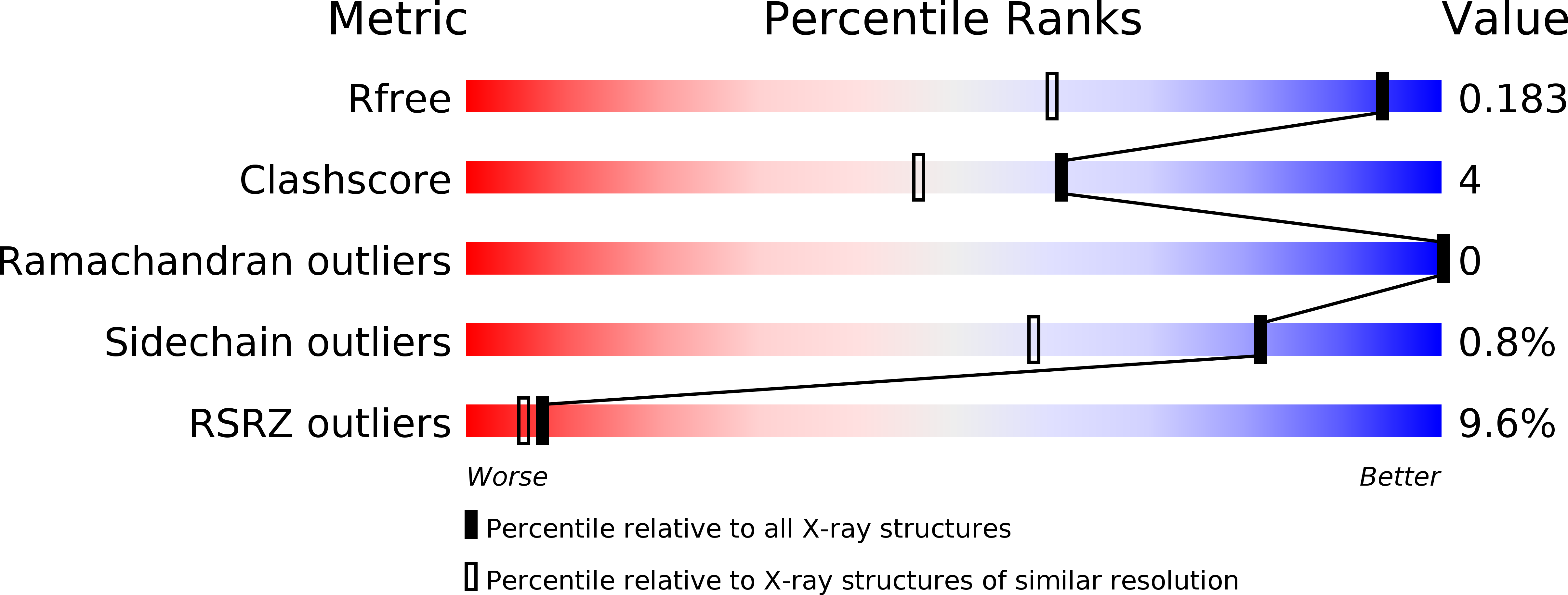
Resolution:
1.30 Å
R-Value Free:
0.18
R-Value Work:
0.15
R-Value Observed:
0.15
Space Group:
P 1 21 1
Macromolecular Entities
Polymer Type:polypeptide(L)
Molecule:Ferrichrome ABC transporter lipoprotein
Chain IDs:A
Chain Length:296
Number of Molecules:1
Biological Source:Staphylococcus aureus subsp. aureus strain
Primary Citation
The Staphylococcus aureus siderophore receptor HtsA undergoes localized conformational changes to enclose staphyloferrin A in an arginine-rich binding pocket.
J.Biol.Chem. 285 11162 11171 (2010)
PMID: 20147287 DOI: 10.1074/jbc.M109.097865

Abstact

Staphylococcus aureus uses several efficient iron acquisition strategies to overcome iron limitation. Recently, the genetic locus encoding biosynthetic enzymes for the iron chelating molecule, staphyloferrin A (SA), was determined. S. aureus synthesizes and secretes SA into its environment to scavenge iron. The membrane-anchored ATP binding cassette-binding protein, HtsA, receives the ferric-chelate for import into the cell. Recently, we determined the apoHtsA crystal structure, the first siderophore receptor from gram-positive bacteria to be structurally characterized. Herein we present the x-ray crystal structure of the HtsA-ferric-SA complex. HtsA adopts a class III binding protein fold composed of separate N- and C-terminal domains bridged by a single alpha-helix. Recombinant HtsA can efficiently sequester ferric-SA from S. aureus culture supernatants where it is bound within the pocket formed between distinct N- and C-terminal domains. A basic patch composed mainly of six Arg residues contact the negatively charged siderophore, securing it within the pocket. The x-ray crystal structures from two different ligand-bound crystal forms were determined. The structures represent the first structural characterization of an endogenous alpha-hydroxycarboxylate-type siderophore-receptor complex. One structure is in an open form similar to apoHtsA, whereas the other is in a more closed conformation. The conformational change is highlighted by isolated movement of three loops within the C-terminal domain, a domain movement unique to known class III binding protein structures.

Legend

Protein

Chemical

Disease

Primary Citation of related structures