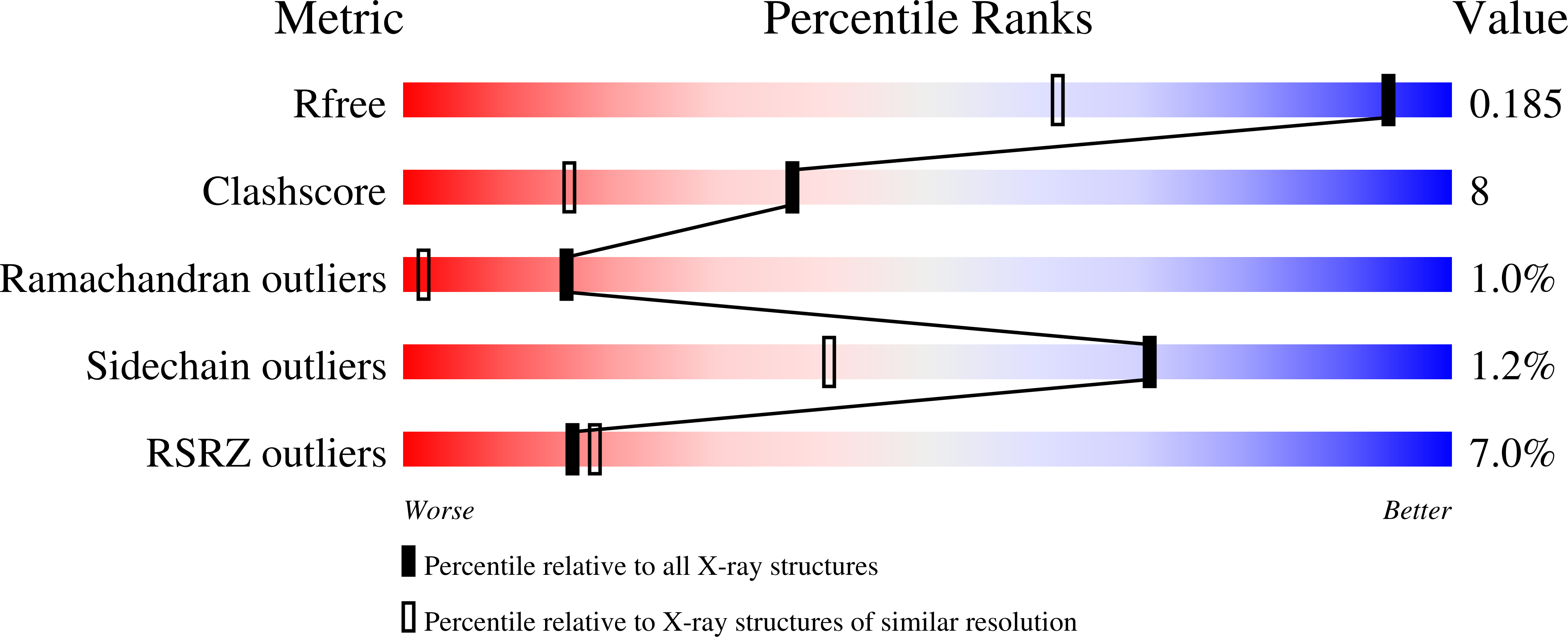
3LHC image
Deposition Date 2010-01-21
Release Date 2010-02-09
Last Version Date 2024-10-09
Entry Detail
PDB ID:
3LHC
Title:
Crystal structure of cyanovirin-n swapping domain b mutant
Biological Source:
Source Organism:
Host Organism:
Method Details:
Experimental Method:
Resolution:
1.34 Å
R-Value Free:
0.22
R-Value Work:
0.19
R-Value Observed:
0.19
Space Group:
P 32 2 1
Macromolecular Entities
Structures with similar UniProt ID
Protein Blast
Polymer Type:polypeptide(L)
Molecule:Cyanovirin-N
Mutagens:E41A,N42A,T57A,R76A,Q78G
Chain IDs:A
Chain Length:100
Number of Molecules:1
Biological Source:Nostoc ellipsosporum
Primary Citation
Anti-HIV activity of defective cyanovirin-N mutants is restored by dimerization.
J.Biol.Chem. 285 13057 13065 (2010)
PMID: 20147291 DOI: 10.1074/jbc.M109.094938

Abstact

Cyanovirin-N (CV-N) is a two-domain, cyanobacterial protein that inhibits human immunodeficiency virus (HIV) at nanomolar concentrations by binding to high mannose sugars on the HIV envelope glycoprotein gp120. The wild type protein can exist as a monomer or a domain-swapped dimer with the monomer and dimer containing two or four sugar binding sites, respectively, one on each domain. Here we demonstrate that monomeric, single binding site mutants are completely inactive and that a single site, whether located on domain A or B, is insufficient to impart the antiviral activity. Linking inactive, monomeric proteins in a head-to-head fashion by an intermolecular disulfide bond or by creating an exclusively domain-swapped dimer via a hinge residue deletion restored antiviral activity to levels similar to that of wild type CV-N. These findings demonstrate unequivocally that multisite binding by CV-N type lectins is necessary for viral inhibition.

Legend

Protein

Chemical

Disease

Primary Citation of related structures