3LGB image
Deposition Date 2010-01-20
Release Date 2010-04-21
Last Version Date 2024-11-13
Entry Detail
PDB ID:
3LGB
Keywords:
Title:
Crystal Structure of the Fe-S Domain of the yeast DNA primase
Biological Source:
Source Organism:
Host Organism:
Method Details:
Experimental Method:
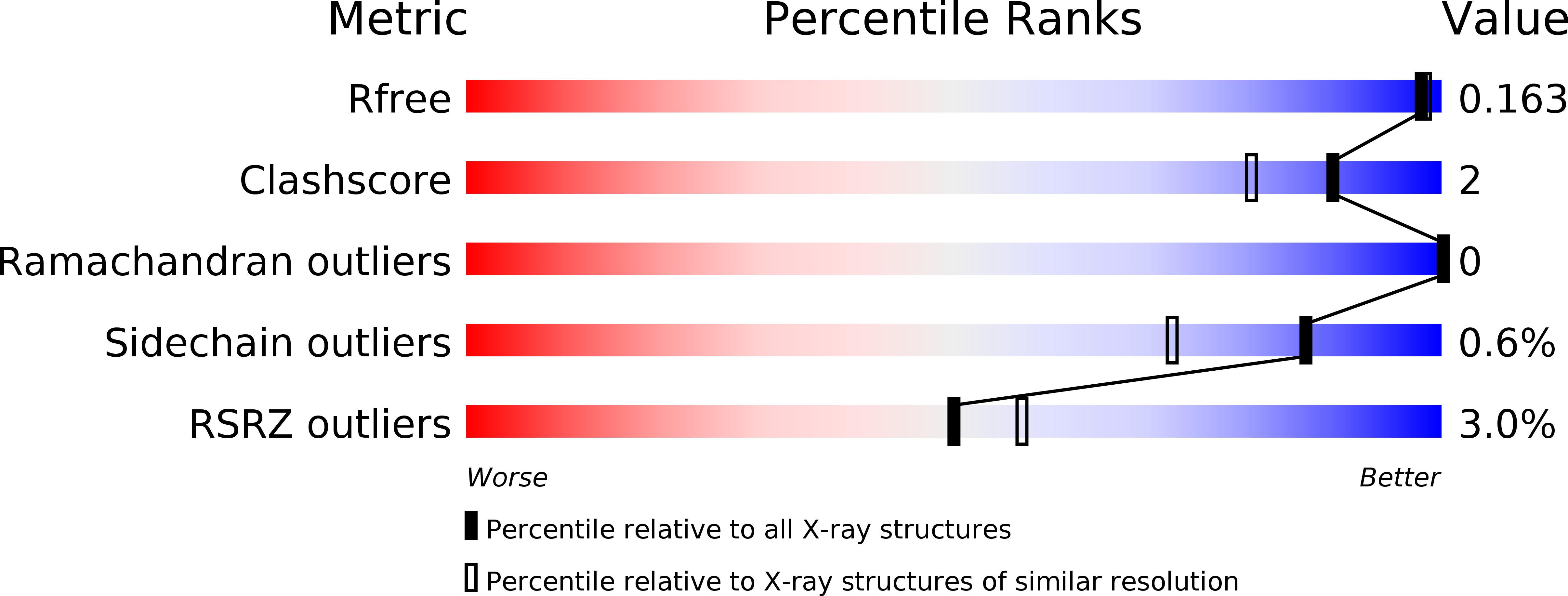
Resolution:
1.54 Å
R-Value Free:
0.16
R-Value Work:
0.15
R-Value Observed:
0.15
Space Group:
P 61
Macromolecular Entities
Polymer Type:polypeptide(L)
Molecule:DNA primase large subunit
Gene (Uniprot):PRI2
Chain IDs:A, B
Chain Length:194
Number of Molecules:2
Biological Source:Saccharomyces cerevisiae
Modified Residue
Compound ID Chain ID Parent Comp ID Details 2D Image
MSE A MET SELENOMETHIONINE
Primary Citation
Shared Active Site Architecture between the Large Subunit of Eukaryotic Primase and DNA Photolyase
PLOS ONE 5 10083 10083 (2010)
PMID: 20404922 DOI: 10.1371/journal.pone.0010083

Abstact

BACKGROUND DNA synthesis during replication relies on RNA primers synthesised by the primase, a specialised DNA-dependent RNA polymerase that can initiate nucleic acid synthesis de novo. In archaeal and eukaryotic organisms, the primase is a heterodimeric enzyme resulting from the constitutive association of a small (PriS) and large (PriL) subunit. The ability of the primase to initiate synthesis of an RNA primer depends on a conserved Fe-S domain at the C-terminus of PriL (PriL-CTD). However, the critical role of the PriL-CTD in the catalytic mechanism of initiation is not understood. METHODOLOGY/PRINCIPAL FINDINGS Here we report the crystal structure of the yeast PriL-CTD at 1.55 A resolution. The structure reveals that the PriL-CTD folds in two largely independent alpha-helical domains joined at their interface by a [4Fe-4S] cluster. The larger N-terminal domain represents the most conserved portion of the PriL-CTD, whereas the smaller C-terminal domain is largely absent in archaeal PriL. Unexpectedly, the N-terminal domain reveals a striking structural similarity with the active site region of the DNA photolyase/cryptochrome family of flavoproteins. The region of similarity includes PriL-CTD residues that are known to be essential for initiation of RNA primer synthesis by the primase. CONCLUSION/SIGNIFICANCE Our study reports the first crystallographic model of the conserved Fe-S domain of the archaeal/eukaryotic primase. The structural comparison with a cryptochrome protein bound to flavin adenine dinucleotide and single-stranded DNA provides important insight into the mechanism of RNA primer synthesis by the primase.

Legend

Protein

Chemical

Disease

Primary Citation of related structures