
Deposition Date
2010-01-18
Release Date
2011-02-02
Last Version Date
2023-09-06
Entry Detail
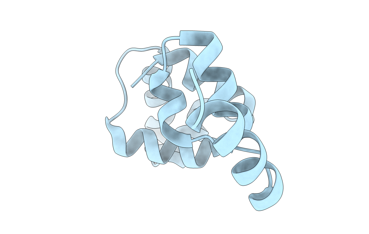
PDB ID:
3LFP
Keywords:
Title:
Crystal Structure of the Restriction-Modification Controller Protein C.Csp231I
Biological Source:
Source Organism(s):
Citrobacter sp. RFL231 (Taxon ID: 315237)
Expression System(s):
Method Details:
Experimental Method:
Resolution:
2.00 Å
R-Value Free:
0.22
R-Value Work:
0.17
R-Value Observed:
0.17
Space Group:
F 41 3 2
