3LFL image
Deposition Date 2010-01-18
Release Date 2010-11-24
Last Version Date 2024-02-21
Entry Detail
PDB ID:
3LFL
Keywords:
Title:
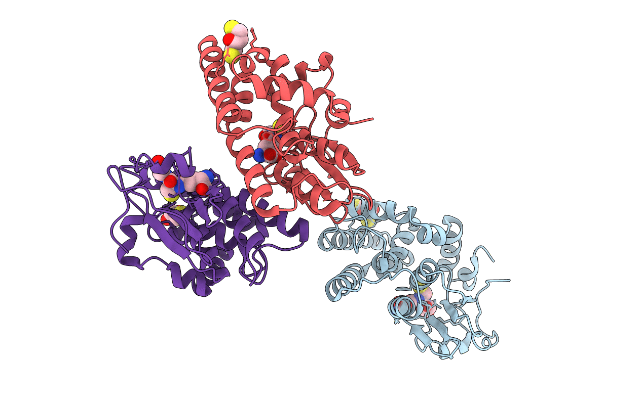
Crystal Structure of human Glutathione Transferase Omega 1, delta 155
Biological Source:
Source Organism:
Homo sapiens (Taxon ID: 9606)
Host Organism:
Method Details:
Experimental Method:
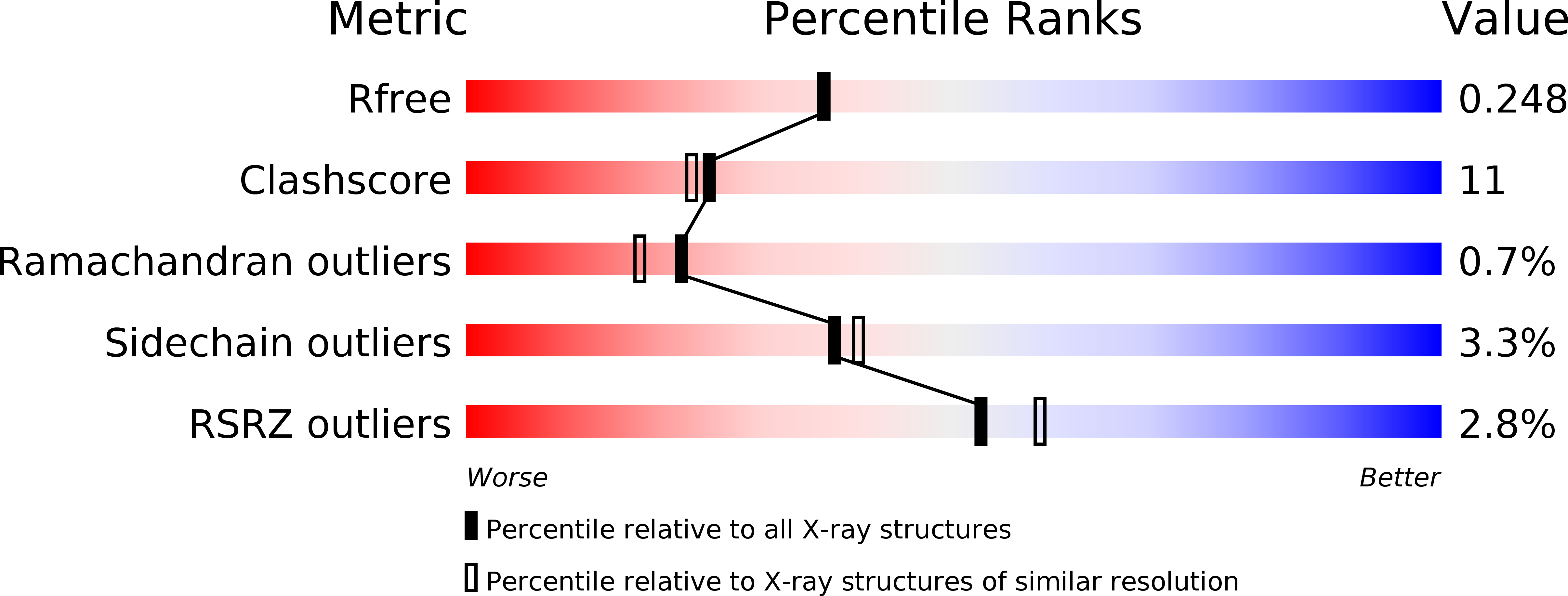
Resolution:
2.10 Å
R-Value Free:
0.25
R-Value Work:
0.19
R-Value Observed:
0.20
Space Group:
P 21 21 2
Macromolecular Entities
Polymer Type:polypeptide(L)
Molecule:Glutathione S-transferase omega-1
Gene (Uniprot):GSTO1
Chain IDs:A, B, C
Chain Length:240
Number of Molecules:3
Biological Source:Homo sapiens
Primary Citation
Novel folding and stability defects cause a deficiency of human glutathione transferase omega 1.
J.Biol.Chem. 286 4271 4279 (2011)
PMID: 21106529 DOI: 10.1074/jbc.M110.197822

Abstact

The polymorphic deletion of Glu-155 from human glutathione transferase omega1 (GSTO1-1) occurs in most populations. Although the recombinant ΔGlu-155 enzyme expressed in Escherichia coli is active, the deletion causes a deficiency of the active enzyme in vivo. The crystal structure and the folding/unfolding kinetics of the ΔGlu-155 variant were determined in order to investigate the cause of the rapid loss of the enzyme in human cells. The crystal structure revealed altered packing around the Glu-155 deletion, an increase in the predicted solvent-accessible area and a corresponding reduction in the buried surface area. This increase in solvent accessibility was consistent with an elevated Stern-Volmer constant. The unfolding of both the wild type and ΔGlu-155 enzyme in urea is best described by a three-state model, and there is evidence for the more pronounced population of an intermediate state by the ΔGlu-155 enzymes. Studies using intrinsic fluorescence revealed a free energy change around 14.4 kcal/mol for the wild type compared with around 8.6 kcal/mol for the ΔGlu-155 variant, which indicates a decrease in stability associated with the Glu-155 deletion. Urea induced unfolding of the wild type GSTO1-1 was reversible through an initial fast phase followed by a second slow phase. In contrast, the ΔGlu-155 variant lacks the slow phase, indicating a refolding defect. It is possible that in some conditions in vivo, the increased solvent-accessible area and the low stability of the ΔGlu-155 variant may promote its unfolding, whereas the refolding defect limits its refolding, resulting in GSTO1-1 deficiency.

Legend

Protein

Chemical

Disease

Primary Citation of related structures