Deposition Date
2010-01-15
Release Date
2010-07-07
Last Version Date
2023-09-06
Entry Detail
PDB ID:
3LF0
Keywords:
Title:
Crystal structure of the ATP bound Mycobacterium tuberculosis nitrogen regulatory PII protein
Biological Source:
Source Organism(s):
Mycobacterium tuberculosis (Taxon ID: 1773)
Expression System(s):
Method Details:
Experimental Method:
Resolution:
2.40 Å
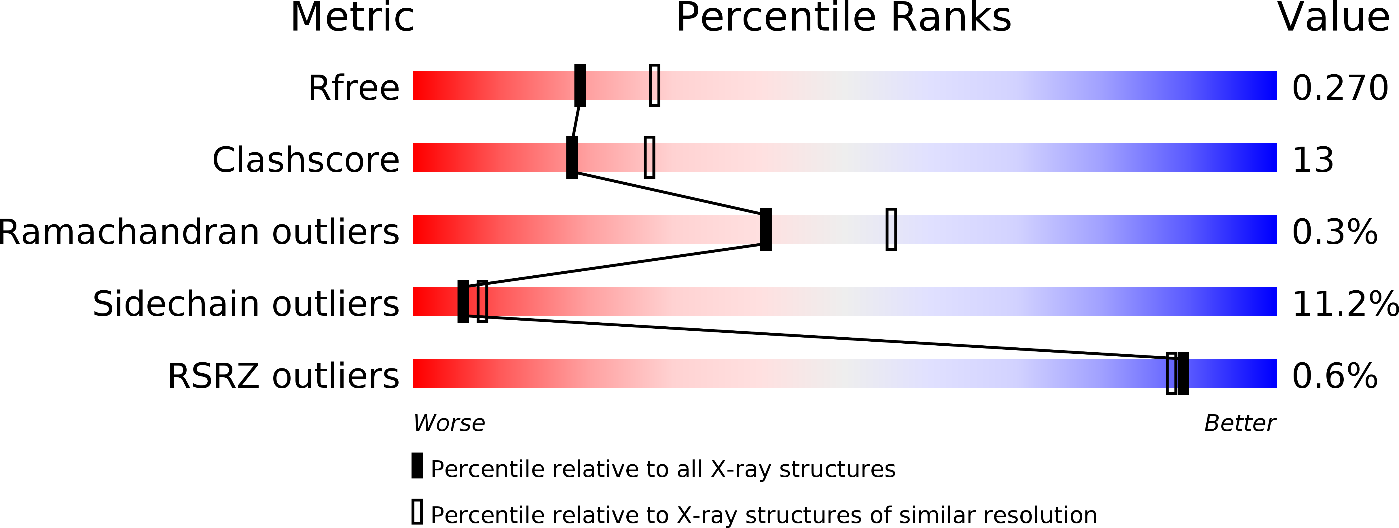
R-Value Free:
0.27
R-Value Work:
0.20
R-Value Observed:
0.20
Space Group:
P 43 21 2