Deposition Date
2010-01-12
Release Date
2010-08-04
Last Version Date
2024-10-09
Entry Detail
PDB ID:
3LDB
Keywords:
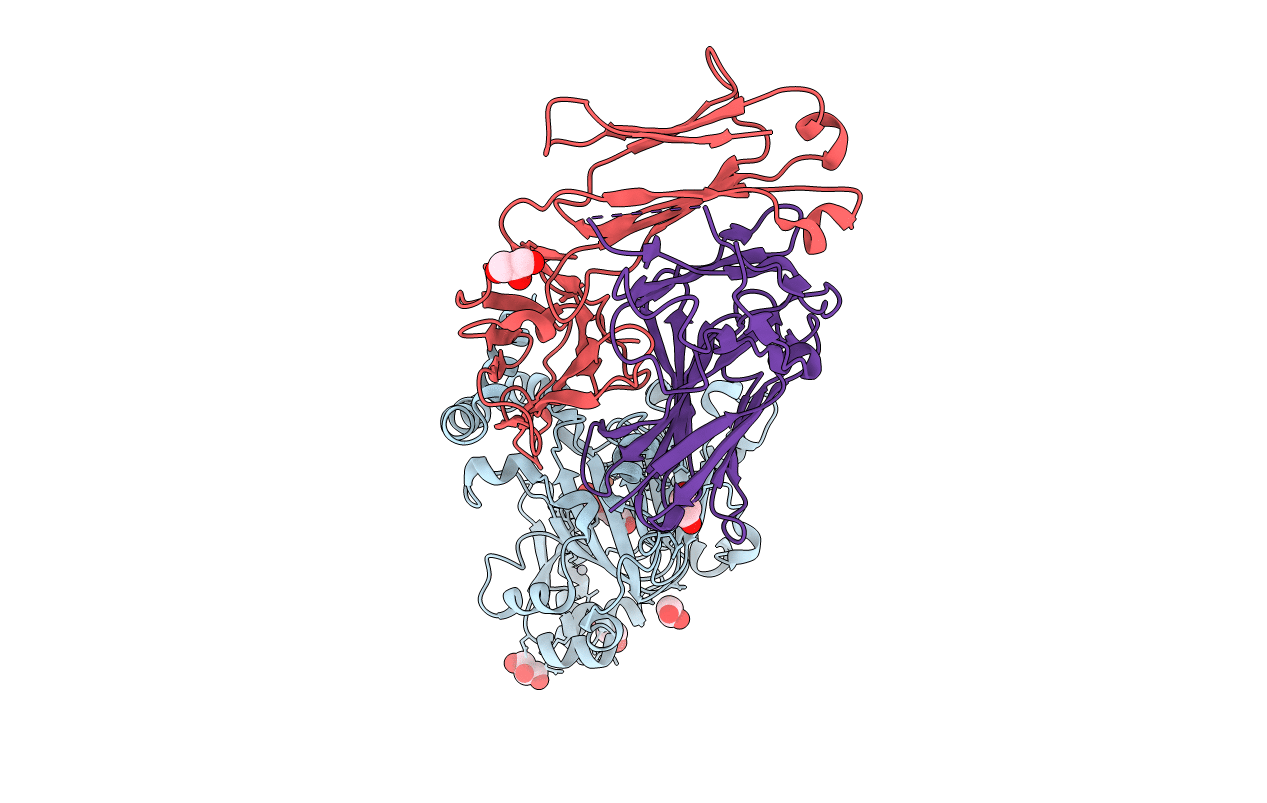
Title:
Structure of JMJD6 complexd with ALPHA-KETOGLUTARATE and Fab Fragment.
Biological Source:
Source Organism(s):
Homo sapiens (Taxon ID: 9606)
Cricetulus migratorius (Taxon ID: 10032)
Cricetulus migratorius (Taxon ID: 10032)
Expression System(s):
Method Details:
Experimental Method:
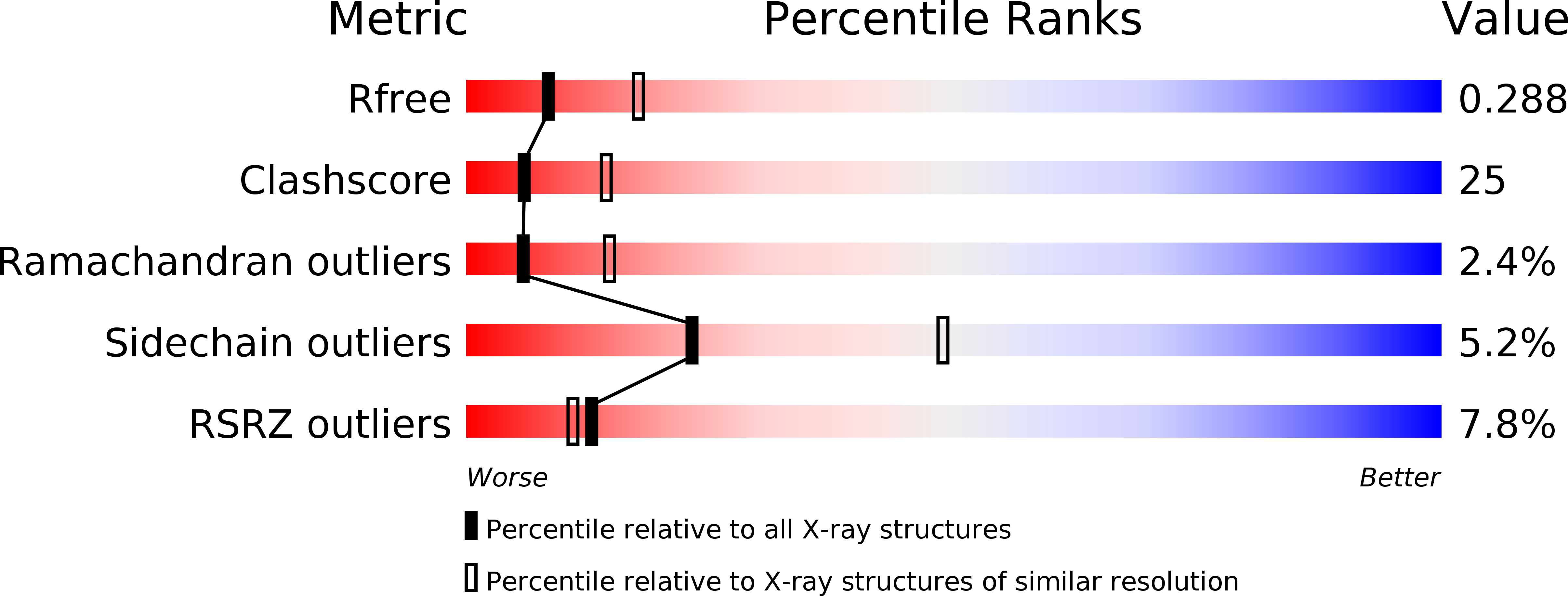
Resolution:
2.70 Å
R-Value Free:
0.28
R-Value Work:
0.25
R-Value Observed:
0.25
Space Group:
P 41 21 2