3LCT image
Deposition Date 2010-01-11
Release Date 2010-08-04
Last Version Date 2023-09-06
Entry Detail
PDB ID:
3LCT
Keywords:
Title:
Crystal Structure of the Anaplastic Lymphoma Kinase Catalytic Domain
Biological Source:
Source Organism:
Homo sapiens (Taxon ID: 9606)
Host Organism:
Method Details:
Experimental Method:
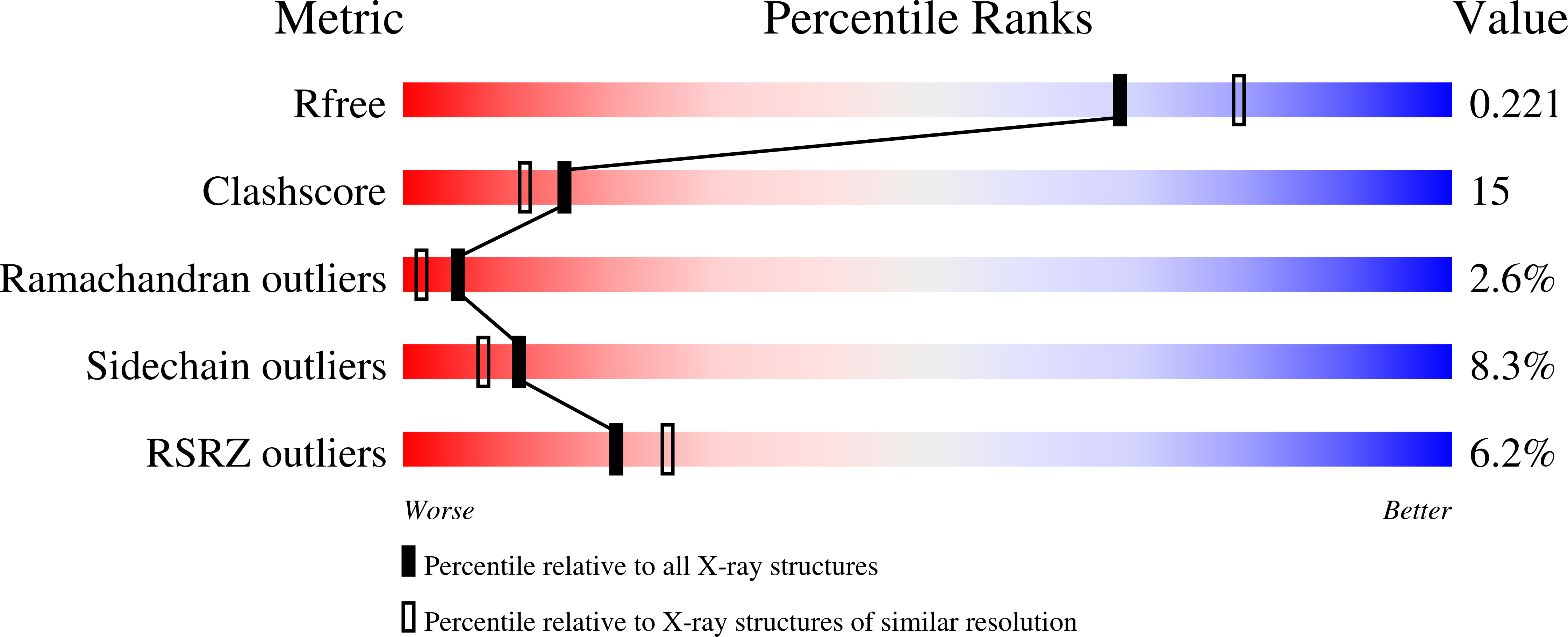
Resolution:
2.10 Å
R-Value Free:
0.25
R-Value Work:
0.20
R-Value Observed:
0.20
Space Group:
P 21 21 21
Macromolecular Entities
Structures with similar UniProt ID
Protein Blast
Polymer Type:polypeptide(L)
Molecule:ALK tyrosine kinase receptor
Gene (Uniprot):ALK
Mutagens:S1281G
Chain IDs:A
Chain Length:344
Number of Molecules:1
Biological Source:Homo sapiens
Primary Citation
Crystal structure of the ALK (anaplastic lymphoma kinase) catalytic domain.
Biochem.J. 430 425 437 (2010)
PMID: 20632993 DOI: 10.1042/BJ20100609

Abstact

ALK (anaplastic lymphoma kinase) is an RTK (receptor tyrosine kinase) of the IRK (insulin receptor kinase) superfamily, which share an YXXXYY autophosphorylation motif within their A-loops (activation loops). A common activation and regulatory mechanism is believed to exist for members of this superfamily typified by IRK and IGF1RK (insulin-like growth factor receptor kinase-1). Chromosomal translocations involving ALK were first identified in anaplastic large-cell lymphoma, a subtype of non-Hodgkin's lymphoma, where aberrant fusion of the ALK kinase domain with the NPM (nucleophosmin) dimerization domain results in autophosphosphorylation and ligand-independent activation. Activating mutations within the full-length ALK kinase domain, most commonly R1275Q and F1174L, which play a major role in neuroblastoma, were recently identified. To provide a structural framework for understanding these mutations and to guide structure-assisted drug discovery efforts, the X-ray crystal structure of the unphosphorylated ALK catalytic domain was determined in the apo, ADP- and staurosporine-bound forms. The structures reveal a partially inactive protein kinase conformation distinct from, and lacking, many of the negative regulatory features observed in inactive IGF1RK/IRK structures in their unphosphorylated forms. The A-loop adopts an inhibitory pose where a short proximal A-loop helix (alphaAL) packs against the alphaC helix and a novel N-terminal beta-turn motif, whereas the distal portion obstructs part of the predicted peptide-binding region. The structure helps explain the reported unique peptide substrate specificity and the importance of phosphorylation of the first A-loop Tyr1278 for kinase activity and NPM-ALK transforming potential. A single amino acid difference in the ALK substrate peptide binding P-1 site (where the P-site is the phosphoacceptor site) was identified that, in conjunction with A-loop sequence variation including the RAS (Arg-Ala-Ser)-motif, rationalizes the difference in the A-loop tyrosine autophosphorylation preference between ALK and IGF1RK/IRK. Enzymatic analysis of recombinant R1275Q and F1174L ALK mutant catalytic domains confirms the enhanced activity and transforming potential of these mutants. The transforming ability of the full-length ALK mutants in soft agar colony growth assays corroborates these findings. The availability of a three-dimensional structure for ALK will facilitate future structure-function and rational drug design efforts targeting this receptor tyrosine kinase.

Legend

Protein

Chemical

Disease

Primary Citation of related structures
Feedback Form
Name
Email
Institute
Feedback