Deposition Date
1997-04-08
Release Date
1997-12-03
Last Version Date
2024-10-30
Entry Detail
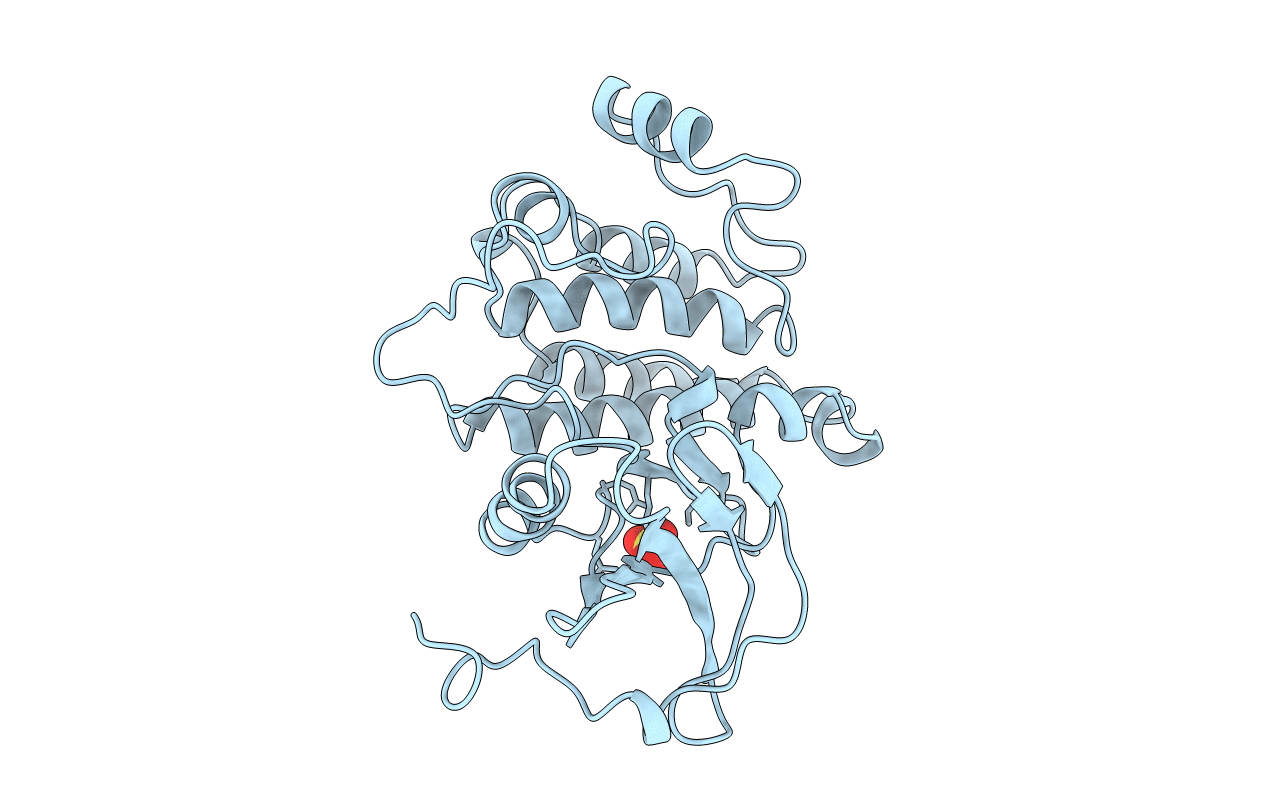
PDB ID:
3LCK
Keywords:
Title:
THE KINASE DOMAIN OF HUMAN LYMPHOCYTE KINASE (LCK), ACTIVATED FORM (AUTO-PHOSPHORYLATED ON TYR394)
Biological Source:
Source Organism(s):
Homo sapiens (Taxon ID: 9606)
Expression System(s):
Method Details:
Experimental Method:
Resolution:
1.70 Å
R-Value Free:
0.19
R-Value Work:
0.17
R-Value Observed:
0.17
Space Group:
P 21 21 21