3LBK image
Deposition Date 2010-01-08
Release Date 2010-03-16
Last Version Date 2023-11-01
Entry Detail
PDB ID:
3LBK
Keywords:
Title:
Structure of human MDM2 protein in complex with a small molecule inhibitor
Biological Source:
Source Organism:
Homo sapiens (Taxon ID: 9606)
Host Organism:
Method Details:
Experimental Method:
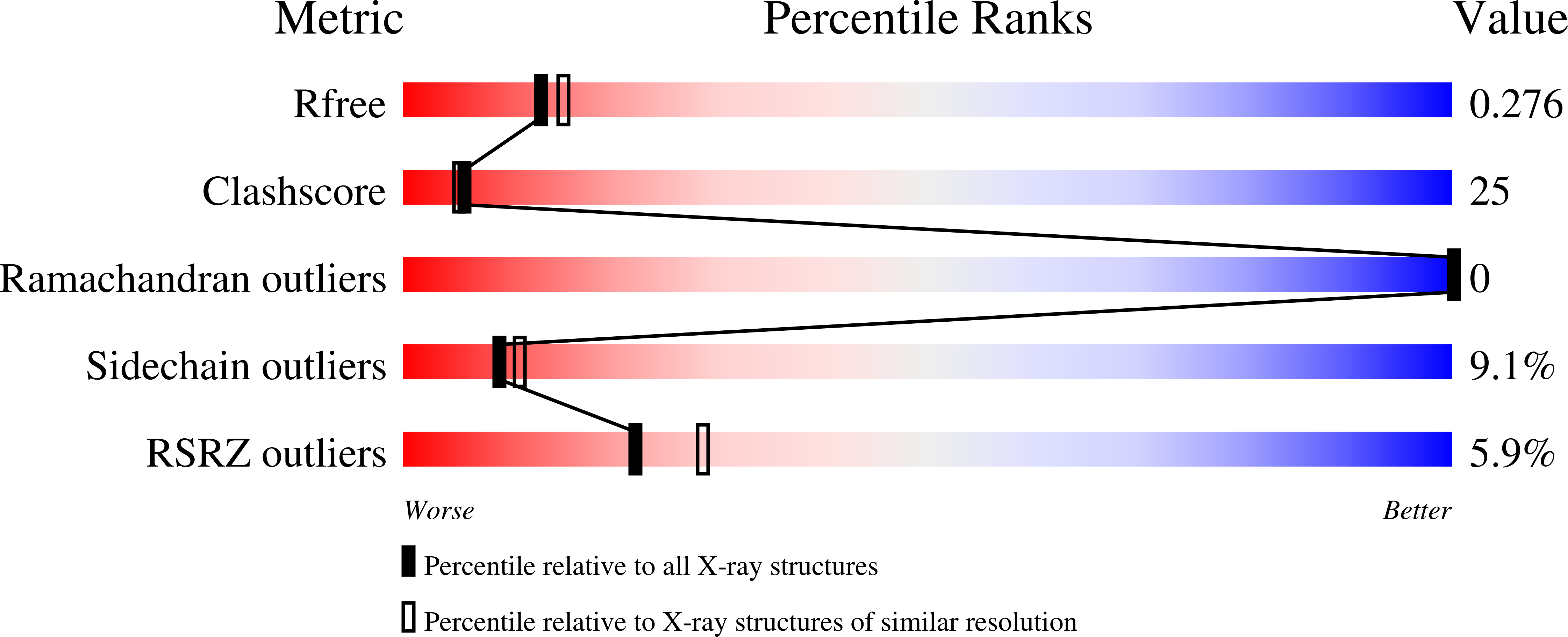
Resolution:
2.30 Å
R-Value Free:
0.25
R-Value Work:
0.20
R-Value Observed:
0.20
Space Group:
P 41 2 2
Macromolecular Entities
Structures with similar UniProt ID
Protein Blast
Polymer Type:polypeptide(L)
Molecule:E3 ubiquitin-protein ligase Mdm2
Gene (Uniprot):MDM2
Mutagens:L33E
Chain IDs:A
Chain Length:95
Number of Molecules:1
Biological Source:Homo sapiens
Primary Citation
Structures of low molecular weight inhibitors bound to MDMX and MDM2 reveal new approaches for p53-MDMX/MDM2 antagonist drug discovery
Cell Cycle 9 1104 1111 (2010)
PMID: 20237429 DOI: 10.4161/cc.9.6.10956

Abstact

Intensive anticancer drug discovery efforts have been made to develop small molecule inhibitors of the p53-MDM2 and p53-MDMX interactions. We present here the structures of the most potent inhibitors bound to MDM2 and MDMX that are based on the new imidazo-indole scaffold. In addition, the structure of the recently reported spiro-oxindole inhibitor bound to MDM2 is described. The structures indicate how the substituents of a small molecule that bind to the three subpockets of the MDM2/X-p53 interaction should be optimized for effective binding to MDM2 and/or MDMX. While the spiro-oxindole inhibitor triggers significant ligand-induced changes in MDM2, the imidazo-indoles share similar binding modes for MDMX and MDM2, but cause only minimal induced-fit changes in the structures of both proteins. Our study includes the first structure of the complex between MDMX and a small molecule and should aid in developing efficient scaffolds for binding to MDMX and/or MDM2.

Legend

Protein

Chemical

Disease

Primary Citation of related structures