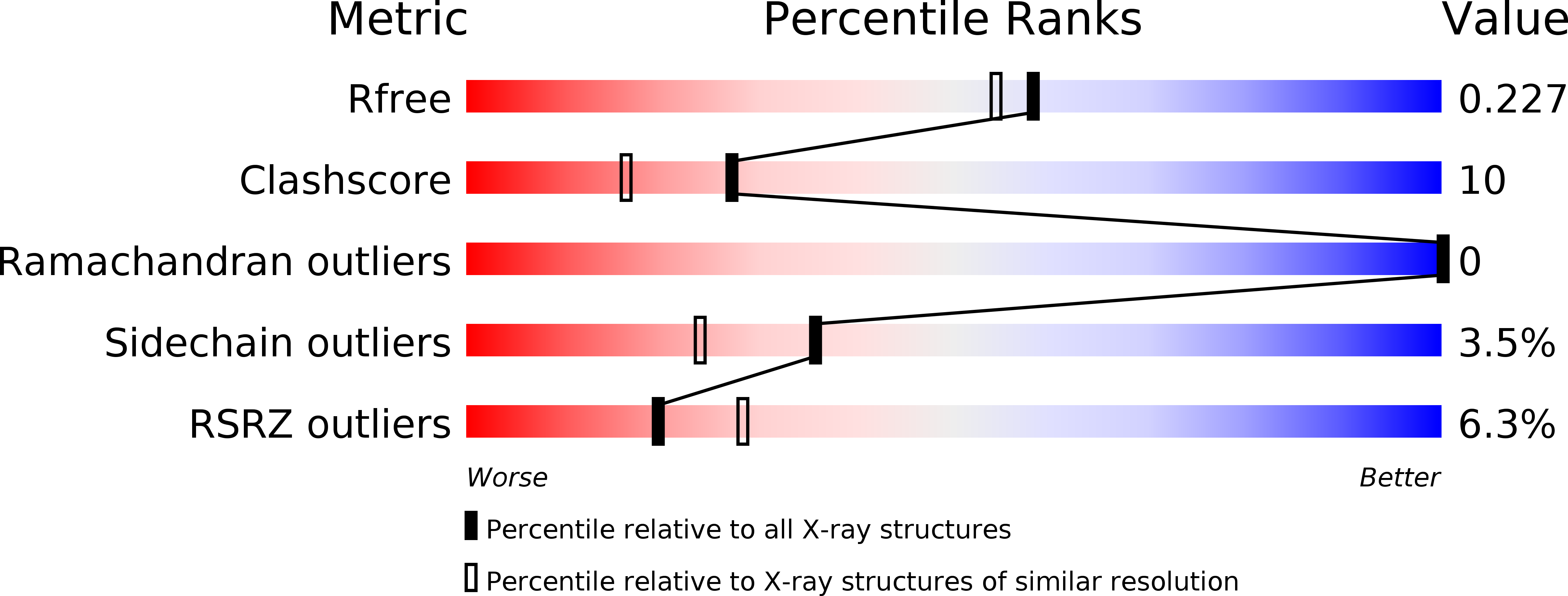
3L9B image
Deposition Date 2010-01-04
Release Date 2011-01-19
Last Version Date 2023-11-01
Entry Detail
PDB ID:
3L9B
Title:
Crystal Structure of Rat Otoferlin C2A
Biological Source:
Source Organism:
Host Organism:
Method Details:
Experimental Method:
Resolution:
1.95 Å
R-Value Free:
0.22
R-Value Work:
0.18
R-Value Observed:
0.18
Space Group:
I 41
Macromolecular Entities
Polymer Type:polypeptide(L)
Molecule:Otoferlin
Gene (Uniprot):Otof
Chain IDs:A
Chain Length:144
Number of Molecules:1
Biological Source:Rattus norvegicus
Ligand Molecules
Primary Citation
The crystal structure of the C2A domain of otoferlin reveals an unconventional top loop region.
J.Mol.Biol. 406 479 490 (2011)
PMID: 21216247 DOI: 10.1016/j.jmb.2010.12.031

Abstact

Otoferlin (Otof), whose genetic mutations cause profound deafness in humans, is a protein composed of at least six C(2) domains, which are known as Ca(2)(+)-binding and phospholipid-binding regions. Mammalian ferlin proteins are proposed to act in membrane fusion events, with Otof being specifically required for exocytosis in auditory hair cells. Ferlin C(2) domains exhibit a rather low level of sequence similarity to those of synaptotagmins, protein kinase C isoforms, or phospholipases. Here, we report the crystal structure of the N-terminal C(2) domain of Otof (C₂A) at 1.95-Å resolution. In contrast to previous predictions, we found that this C(2) domain is complete with eight β-strands. Comparing the structure of Otof C₂A to those of other C(2) domains revealed one top loop in Otof to be significantly shorter. This results in a depression of the surface, which is positively charged for the Otof C₂A domain, and contrasts with the head-like protrusion surrounded by a negatively charged "neck" typically found in other C(2) domains. Isothermal titration calorimetry and circular dichroism spectroscopy studies confirmed that Otof C₂A is unable to bind Ca(2+), while the synaptotagmin-1 C₂A domain exhibited Ca(2+) binding under the same conditions. Furthermore, floatation assays revealed a failure of Otof C(2)A to bind to phospholipid membranes. Accordingly, no positively charged β-groove-like surface structure, which is known to bind phosphatidylinositol-4,5-bisphosphate in other C(2) domains, was found at the respective position in Otof C₂A. Taken together, these data demonstrate that the Otof C₂A domain differs structurally and functionally from other C(2) domains.

Legend

Protein

Chemical

Disease

Primary Citation of related structures