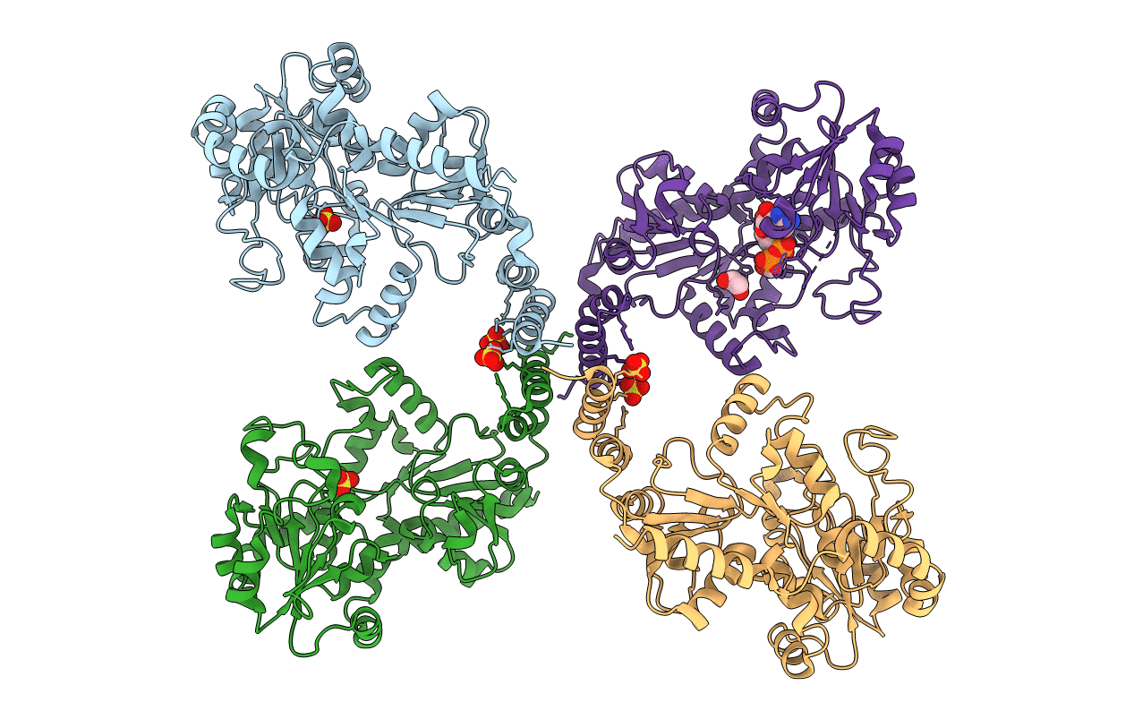
3L7L image
Entry Detail
PDB ID:
3L7L
Title:
Structure of the Wall Teichoic Acid Polymerase TagF, H444N + CDPG (30 minute soak)
Biological Source:
Host Organism:
PDB Version:
Deposition Date:
2009-12-28
Release Date:
2010-04-28
Method Details:
Experimental Method:
Resolution:
2.95 Å
R-Value Free:
0.25
R-Value Work:
0.19
R-Value Observed:
0.19
Space Group:
P 41 21 2
Macromolecular Entities
Polymer Type:polypeptide(L)
Description:Teichoic acid biosynthesis protein F
Mutations:H444N
Chain IDs:A, B, C, D
Chain Length:729
Number of Molecules:4
Biological Source:Staphylococcus epidermidis
Primary Citation
Structure of the bacterial teichoic acid polymerase TagF provides insights into membrane association and catalysis.
Nat.Struct.Mol.Biol. 17 582 589 (2010)
PMID: 20400947 DOI: 10.1038/nsmb.1819

Abstact

Teichoic acid polymers are composed of polyol-phosphate units and form a major component of Gram-positive bacterial cell walls. These anionic compounds perform a multitude of important roles in bacteria and are synthesized by monotopic membrane proteins of the TagF polymerase family. We have determined the structure of Staphylococcus epidermidis TagF to 2.7-A resolution from a construct that includes both the membrane-targeting region and the glycerol-phosphate polymerase domains. TagF possesses a helical region for interaction with the lipid bilayer, placing the active site at a suitable distance for access to the membrane-bound substrate. Characterization of active-site residue variants and analysis of a CDP-glycerol substrate complex suggest a mechanism for polymer synthesis. With the importance of teichoic acid in Gram-positive physiology, this elucidation of the molecular details of TagF function provides a critical new target in the development of novel anti-infectives.

Legend

Protein

Chemical

Disease

Primary Citation of related structures