Deposition Date
2009-12-23
Release Date
2010-01-26
Last Version Date
2024-04-03
Entry Detail
PDB ID:
3L6C
Keywords:
Title:
X-ray crystal structure of rat serine racemase in complex with malonate a potent inhibitor
Biological Source:
Source Organism(s):
Rattus norvegicus (Taxon ID: 10116)
Expression System(s):
Method Details:
Experimental Method:
Resolution:
2.20 Å
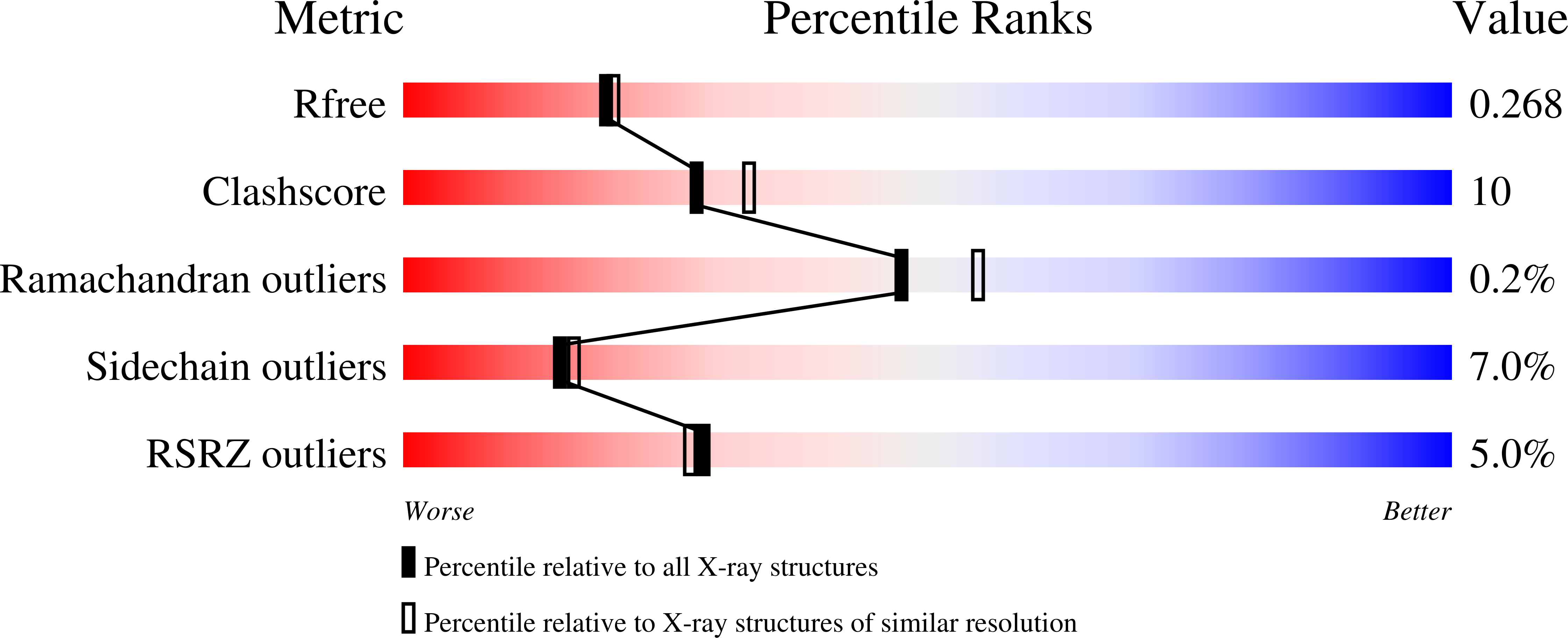
R-Value Free:
0.26
R-Value Work:
0.21
R-Value Observed:
0.21
Space Group:
P 3