3L5V image
Deposition Date 2009-12-22
Release Date 2010-03-09
Last Version Date 2023-09-06
Entry Detail
PDB ID:
3L5V
Keywords:
Title:
Crystal structure of macrophage migration inhibitory factor (MIF) with glycerol at 1.70A resolution
Biological Source:
Source Organism:
Homo sapiens (Taxon ID: 9606)
Host Organism:
Method Details:
Experimental Method:
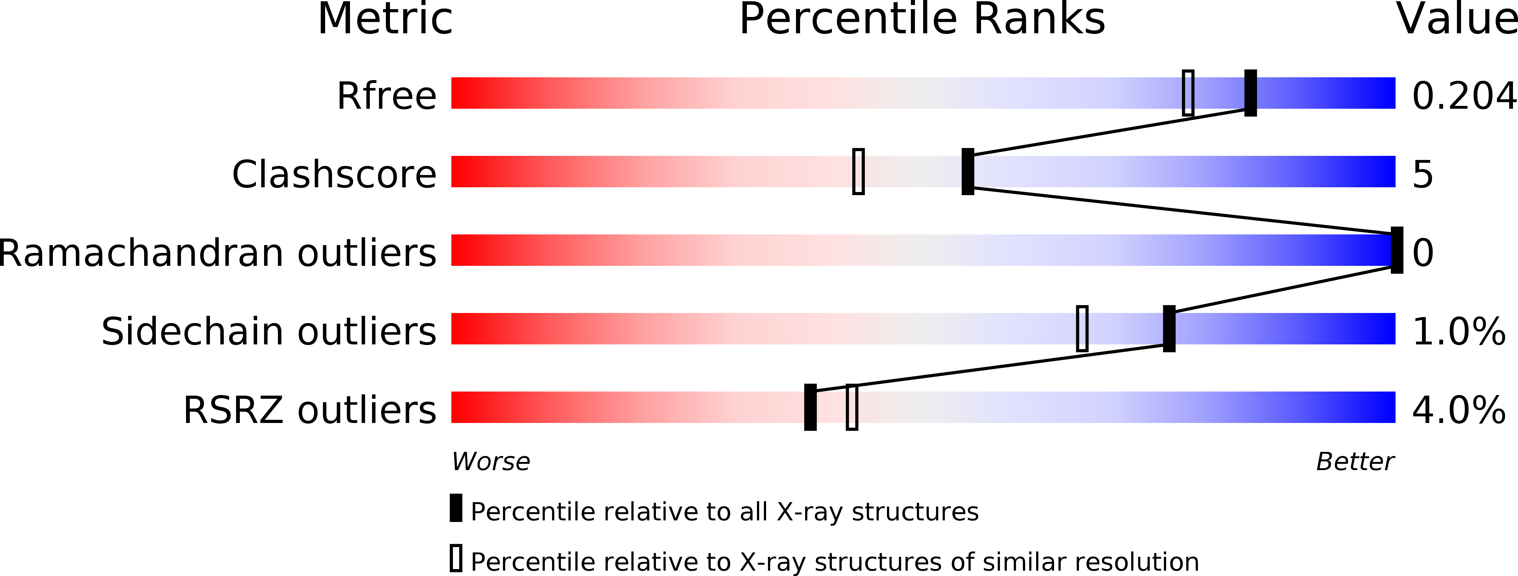
Resolution:
1.70 Å
R-Value Free:
0.21
R-Value Work:
0.19
R-Value Observed:
0.19
Space Group:
P 21 21 21
Macromolecular Entities
Polymer Type:polypeptide(L)
Molecule:Macrophage migration inhibitory factor
Gene (Uniprot):MIF
Chain IDs:A, B, C
Chain Length:122
Number of Molecules:3
Biological Source:Homo sapiens
Primary Citation
Fragment screening of inhibitors for MIF tautomerase reveals a cryptic surface binding site.
Bioorg.Med.Chem.Lett. 20 1821 1824 (2010)
PMID: 20185308 DOI: 10.1016/j.bmcl.2010.02.009

Abstact

In the course of a fragment screening campaign by in silico docking followed by X-ray crystallography, a novel binding site for migration inhibitory factor (MIF) inhibitors was demonstrated. The site is formed by rotation of the side-chain of Tyr-36 to reveal a surface binding site in MIF that is hydrophobic and surrounded by aromatic side-chain residues. The crystal structures of two small inhibitors that bind to this site and of a quinolinone inhibitor, that spans the canonical deep pocket near Pro-1 and the new surface binding site, have been solved. These results suggest new opportunities for structure-based design of MIF inhibitors.

Legend

Protein

Chemical

Disease

Primary Citation of related structures