3L56 image
Deposition Date 2009-12-21
Release Date 2010-03-09
Last Version Date 2023-09-06
Entry Detail
PDB ID:
3L56
Keywords:
Title:
Crystal structure of the large c-terminal domain of polymerase basic protein 2 from influenza virus a/viet nam/1203/2004 (h5n1)
Biological Source:
Source Organism:
Host Organism:
Method Details:
Experimental Method:
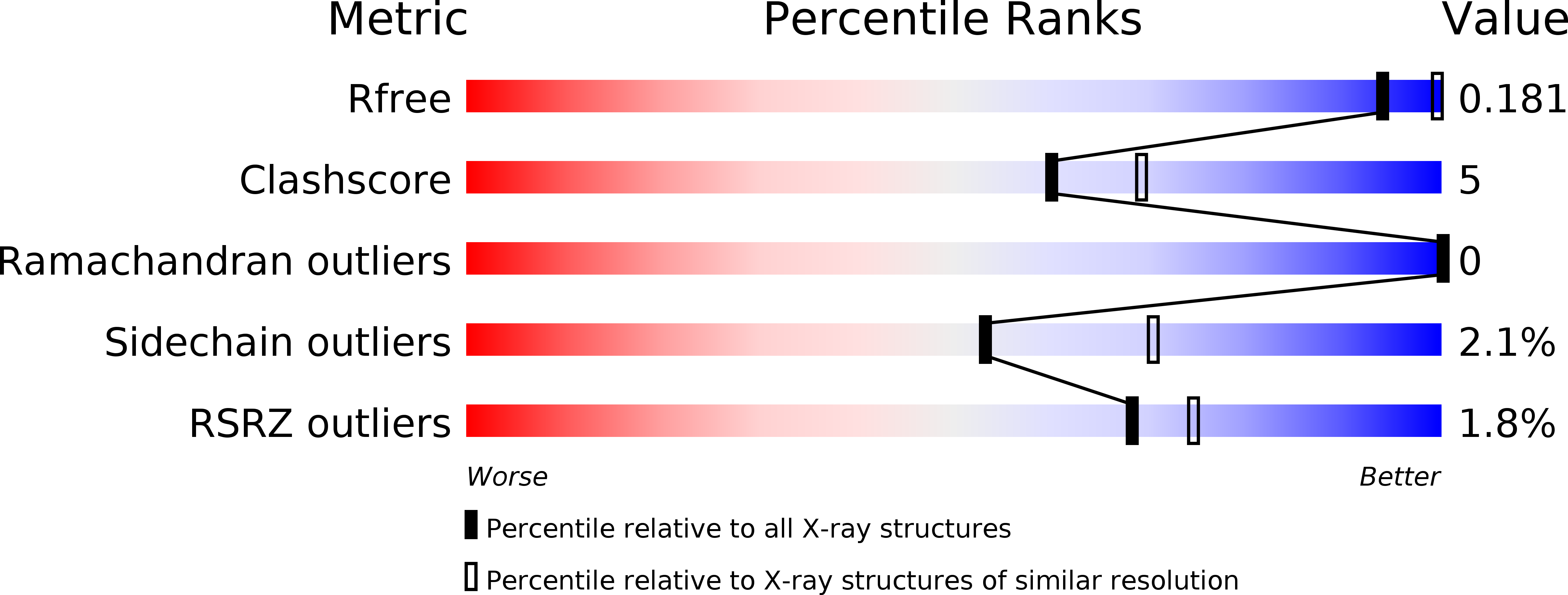
Resolution:
2.30 Å
R-Value Free:
0.23
R-Value Work:
0.17
R-Value Observed:
0.18
Space Group:
P 1 21 1
Macromolecular Entities
Polymer Type:polypeptide(L)
Molecule:Polymerase PB2
Gene (Uniprot):PB2
Chain IDs:A, B
Chain Length:222
Number of Molecules:2
Biological Source:Influenza A virus
Primary Citation
Biological and structural characterization of a host-adapting amino acid in influenza virus.
Plos Pathog. 6 e1001034 e1001034 (2010)
PMID: 20700447 DOI: 10.1371/journal.ppat.1001034

Abstact

Two amino acids (lysine at position 627 or asparagine at position 701) in the polymerase subunit PB2 protein are considered critical for the adaptation of avian influenza A viruses to mammals. However, the recently emerged pandemic H1N1 viruses lack these amino acids. Here, we report that a basic amino acid at position 591 of PB2 can compensate for the lack of lysine at position 627 and confers efficient viral replication to pandemic H1N1 viruses in mammals. Moreover, a basic amino acid at position 591 of PB2 substantially increased the lethality of an avian H5N1 virus in mice. We also present the X-ray crystallographic structure of the C-terminus of a pandemic H1N1 virus PB2 protein. Arginine at position 591 fills the cleft found in H5N1 PB2 proteins in this area, resulting in differences in surface shape and charge for H1N1 PB2 proteins. These differences may affect the protein's interaction with viral and/or cellular factors, and hence its ability to support virus replication in mammals.

Legend

Protein

Chemical

Disease

Primary Citation of related structures