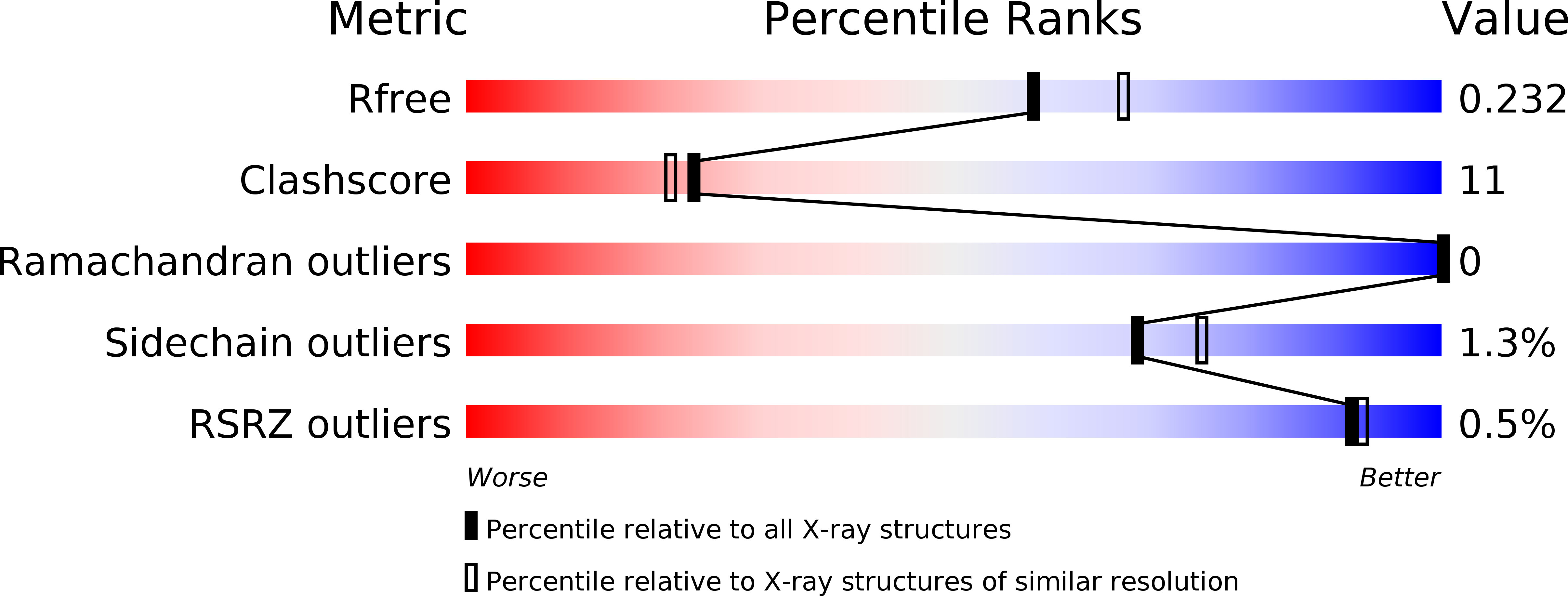
3L48 image
Deposition Date 2009-12-18
Release Date 2010-03-02
Last Version Date 2024-11-06
Entry Detail
PDB ID:
3L48
Title:
Crystal structure of the C-terminal domain of the PapC usher
Biological Source:
Source Organism:
Host Organism:
Method Details:
Experimental Method:
Resolution:
2.10 Å
R-Value Free:
0.22
R-Value Work:
0.19
R-Value Observed:
0.20
Space Group:
P 42 21 2
Macromolecular Entities
Structures with similar UniProt ID
Protein Blast
Polymer Type:polypeptide(L)
Molecule:Outer membrane usher protein PapC
Gene (Uniprot):papC
Chain IDs:A, B, C, D, E
Chain Length:94
Number of Molecules:5
Biological Source:Escherichia coli
Ligand Molecules
Primary Citation
Structural Homology between the C-Terminal Domain of the PapC Usher and Its Plug.
J.Bacteriol. 192 1824 1831 (2010)
PMID: 20118254 DOI: 10.1128/JB.01677-09

Abstact

P pili are extracellular appendages responsible for the targeting of uropathogenic Escherichia coli to the kidney. They are assembled by the chaperone-usher (CU) pathway of pilus biogenesis involving two proteins, the periplasmic chaperone PapD and the outer membrane assembly platform, PapC. Many aspects of the structural biology of the Pap CU pathway have been elucidated, except for the C-terminal domain of the PapC usher, the structure of which is unknown. In this report, we identify a stable and folded fragment of the C-terminal region of the PapC usher and determine its structure using both X-ray crystallography and nuclear magnetic resonance (NMR) spectroscopy. These structures reveal a beta-sandwich fold very similar to that of the plug domain, a domain of PapC obstructing its translocation domain. This structural similarity suggests similar functions in usher-mediated pilus biogenesis, playing out at different stages of the process. This structure paves the way for further functional analysis targeting surfaces common to both the plug and the C-terminal domain of PapC.

Legend

Protein

Chemical

Disease

Primary Citation of related structures