Deposition Date
2009-12-16
Release Date
2010-02-16
Last Version Date
2024-03-20
Entry Detail
PDB ID:
3L32
Keywords:
Title:
Structure of the dimerisation domain of the rabies virus phosphoprotein
Biological Source:
Source Organism(s):
Rabies virus (Taxon ID: 445791)
Expression System(s):
Method Details:
Experimental Method:
Resolution:
1.50 Å
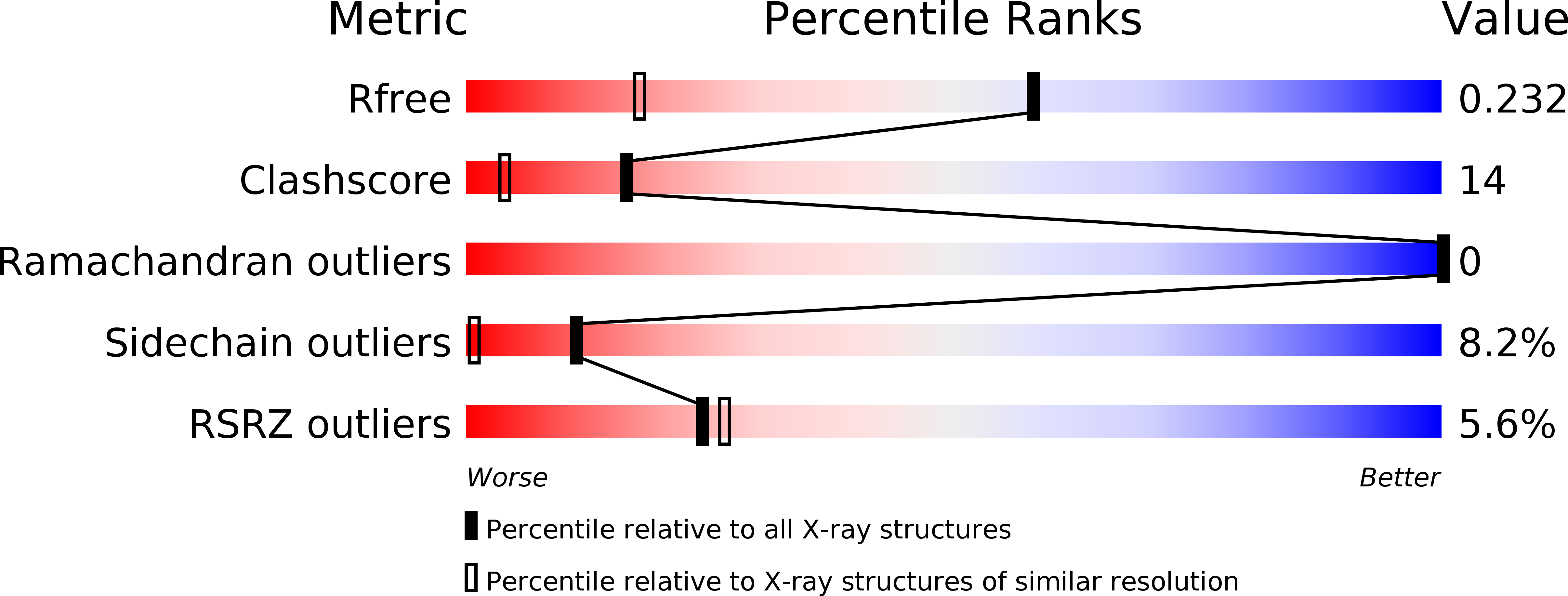
R-Value Free:
0.22
R-Value Work:
0.19
R-Value Observed:
0.19
Space Group:
I 41 2 2