3L2A image
Entry Detail
PDB ID:
3L2A
Title:
Crystal structure of Reston Ebola VP35 interferon inhibitory domain
Biological Source:
Source Organism:
Host Organism:
PDB Version:
Deposition Date:
2009-12-14
Release Date:
2010-05-12
Method Details:
Experimental Method:
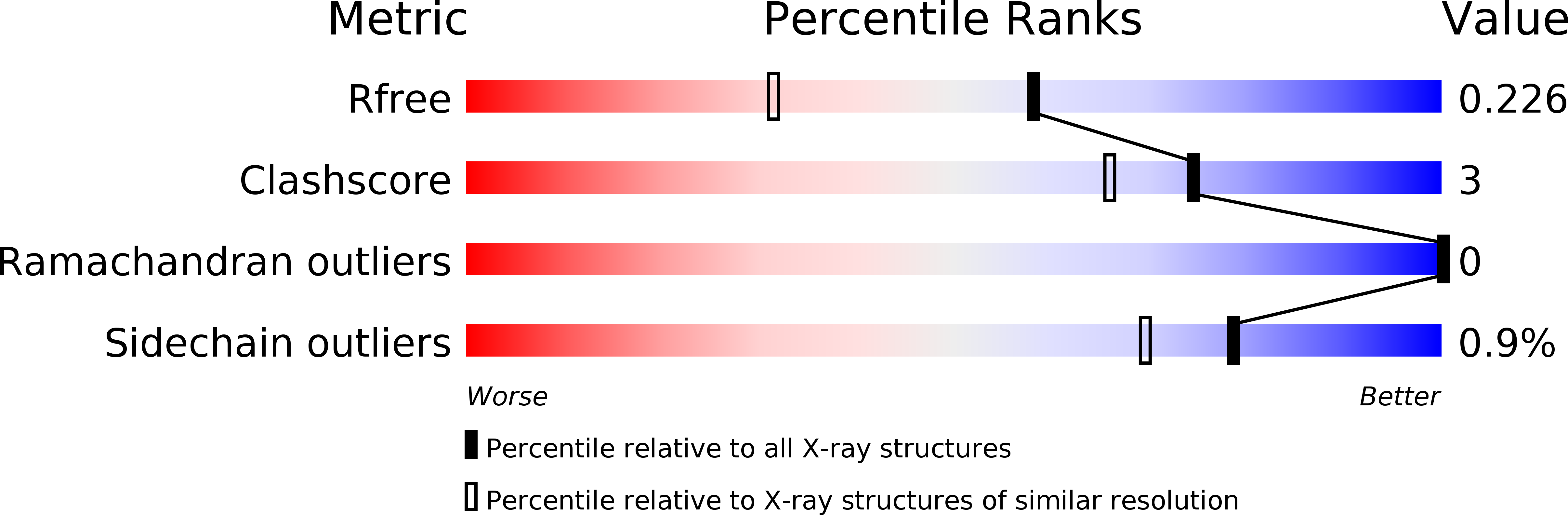
Resolution:
1.71 Å
R-Value Free:
0.22
R-Value Work:
0.17
R-Value Observed:
0.17
Space Group:
P 41
Macromolecular Entities
Polymer Type:polypeptide(L)
Description:Polymerase cofactor VP35
Chain IDs:A
Chain Length:129
Number of Molecules:1
Biological Source:Reston ebolavirus
Primary Citation
Structural and Functional Characterization of Reston Ebola Virus VP35 Interferon Inhibitory Domain.
J.Mol.Biol. 399 347 357 (2010)
PMID: 20399790 DOI: 10.1016/j.jmb.2010.04.022

Abstact

Ebolaviruses are causative agents of lethal hemorrhagic fever in humans and nonhuman primates. Among the filoviruses characterized thus far, Reston Ebola virus (REBOV) is the only Ebola virus that is nonpathogenic to humans despite the fact that REBOV can cause lethal disease in nonhuman primates. Previous studies also suggest that REBOV is less effective at inhibiting host innate immune responses than Zaire Ebola virus (ZEBOV) or Marburg virus. Virally encoded VP35 protein is critical for immune suppression, but an understanding of the relative contributions of VP35 proteins from REBOV and other filoviruses is currently lacking. In order to address this question, we characterized the REBOV VP35 interferon inhibitory domain (IID) using structural, biochemical, and virological studies. These studies reveal differences in double-stranded RNA binding and interferon inhibition between the two species. These observed differences are likely due to increased stability and loss of flexibility in REBOV VP35 IID, as demonstrated by thermal shift stability assays. Consistent with this finding, the 1.71-A crystal structure of REBOV VP35 IID reveals that it is highly similar to that of ZEBOV VP35 IID, with an overall backbone r.m.s.d. of 0.64 A, but contains an additional helical element at the linker between the two subdomains of VP35 IID. Mutations near the linker, including swapping sequences between REBOV and ZEBOV, reveal that the linker sequence has limited tolerance for variability. Together with the previously solved ligand-free and double-stranded-RNA-bound forms of ZEBOV VP35 IID structures, our current studies on REBOV VP35 IID reinforce the importance of VP35 in immune suppression. Functional differences observed between REBOV and ZEBOV VP35 proteins may contribute to observed differences in pathogenicity, but these are unlikely to be the major determinant. However, the high level of similarity in structure and the low tolerance for sequence variability, coupled with the multiple critical roles played by Ebola virus VP35 proteins, highlight the viability of VP35 as a potential target for therapeutic development.

Legend

Protein

Chemical

Disease

Primary Citation of related structures