Deposition Date
2009-12-14
Release Date
2010-05-05
Last Version Date
2023-09-06
Entry Detail
PDB ID:
3L1X
Keywords:
Title:
Crystal Structure of U-box Domain of Human E4B Ubiquitin Ligase
Biological Source:
Source Organism(s):
Homo sapiens (Taxon ID: 9606)
Expression System(s):
Method Details:
Experimental Method:
Resolution:
2.60 Å
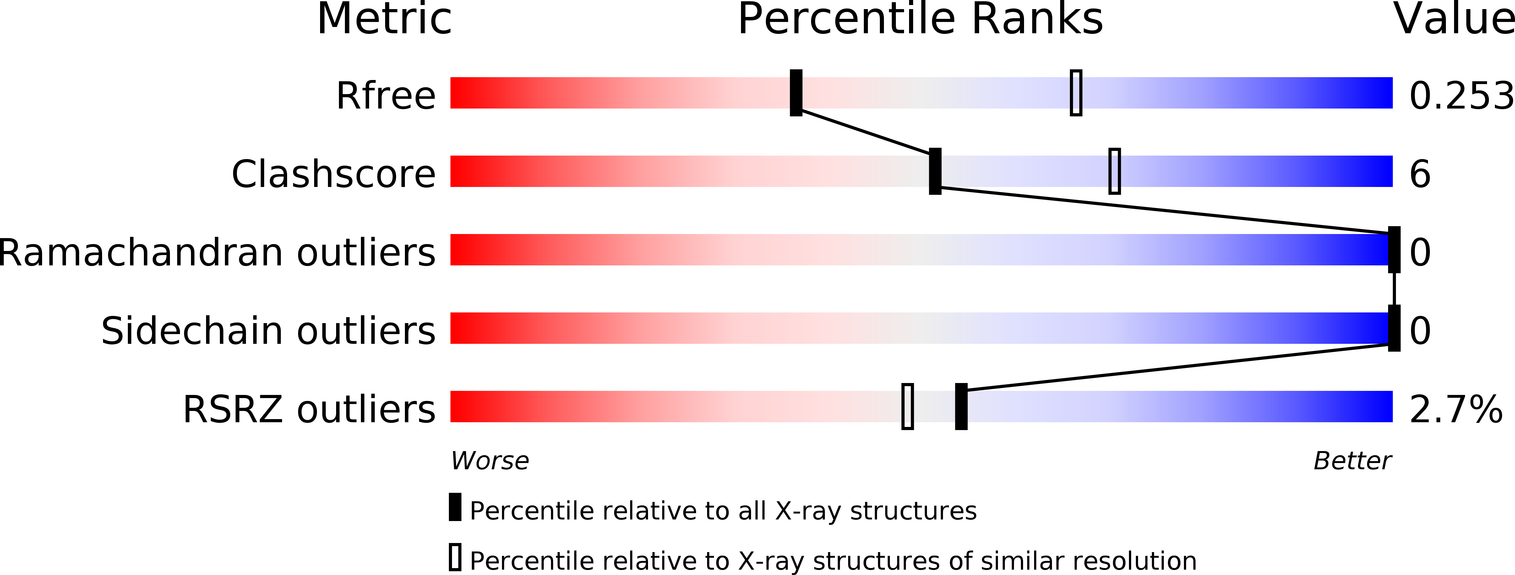
R-Value Free:
0.25
R-Value Work:
0.18
R-Value Observed:
0.19
Space Group:
P 4 3 2