3L1M image
Deposition Date 2009-12-13
Release Date 2010-04-28
Last Version Date 2024-11-06
Entry Detail
PDB ID:
3L1M
Title:
Crystal Structure of a Ni-directed Dimer of Cytochrome cb562 with a Quinolate-Histidine Hybrid Coordination Motif
Biological Source:
Source Organism:
Escherichia coli (Taxon ID: 562)
Method Details:
Experimental Method:
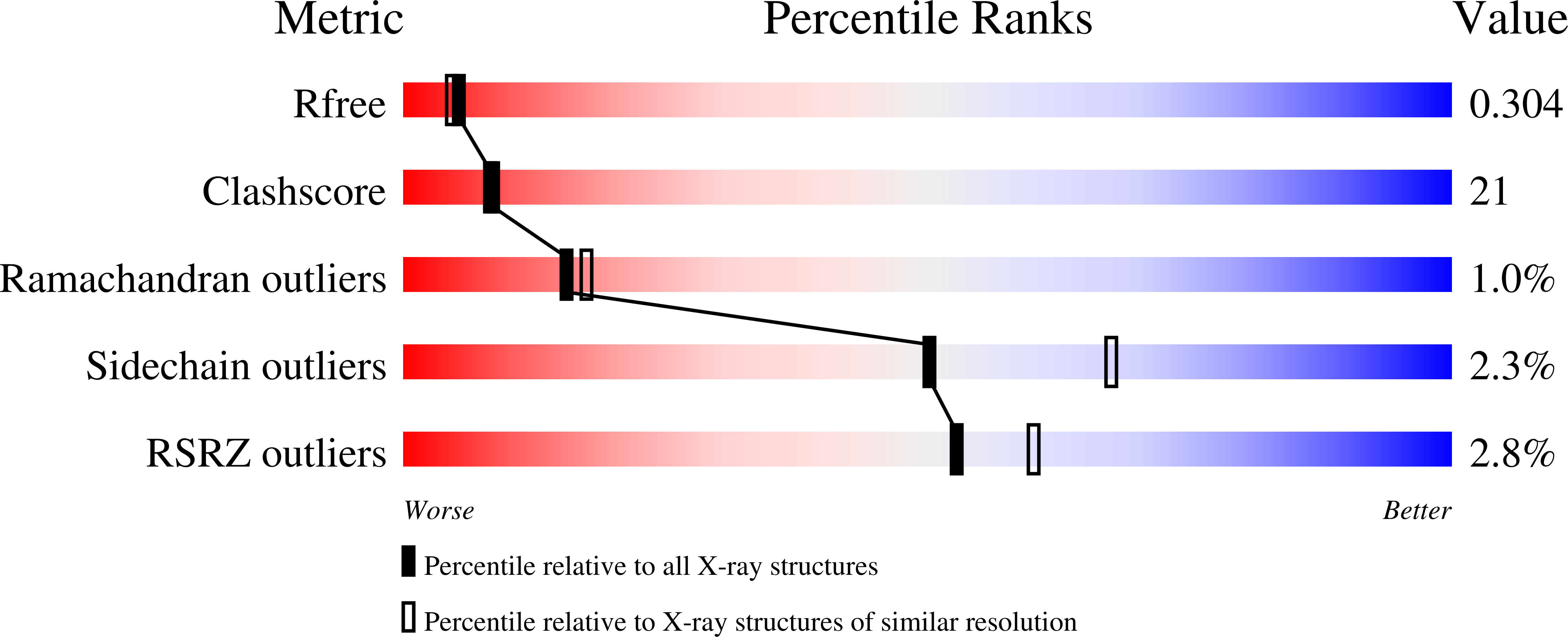
Resolution:
2.30 Å
R-Value Free:
0.31
R-Value Work:
0.26
R-Value Observed:
0.27
Space Group:
P 21 21 2
Macromolecular Entities
Structures with similar UniProt ID
Protein Blast
Polymer Type:polypeptide(L)
Molecule:Soluble cytochrome b562
Gene (Uniprot):cybC
Mutagens:K59W, G70C, R98C, Y101C
Chain IDs:A
Chain Length:106
Number of Molecules:1
Biological Source:Escherichia coli
Primary Citation
Controlled protein dimerization through hybrid coordination motifs.
Inorg.Chem. 49 4362 4369 (2010)
PMID: 20377257 DOI: 10.1021/ic100534y

Abstact

Protein homodimerization is the simplest form of oligomerization that is frequently utilized for the construction of functional biological assemblies and the regulation of cellular pathways. Despite its simplicity, dimerization still poses an enormous challenge for protein engineering and chemical manipulation, owing to the large molecular surfaces involved in this process. We report here the construction of a hybrid coordination motif--consisting of a natural (His) and a non-natural ligand (quinolate)--on the alpha-helical surface of cytochrome cb(562), which (a) simultaneously binds divalent metals with high affinity, (b) leads to a metal-induced increase in global protein stability, and importantly, (c) enables the formation of a discrete protein dimer, whose shape is dictated by the inner-sphere metal coordination geometry and closely approximates that of the DNA-binding domains of bZIP family transcription factors.

Legend

Protein

Chemical

Disease

Primary Citation of related structures