
Deposition Date
2009-12-08
Release Date
2010-03-31
Last Version Date
2023-11-01
Entry Detail
PDB ID:
3KZ8
Keywords:
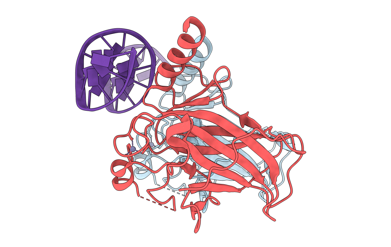
Title:
Diversity in DNA recognition by p53 revealed by crystal structures with Hoogsteen base pairs (p53-DNA complex 3)
Biological Source:
Source Organism(s):
Homo sapiens (Taxon ID: 9606)
Expression System(s):
Method Details:
Experimental Method:
Resolution:
1.91 Å
R-Value Free:
0.26
R-Value Work:
0.22
R-Value Observed:
0.22
Space Group:
C 1 2 1
