3KYQ image
Deposition Date 2009-12-07
Release Date 2010-03-02
Last Version Date 2023-11-01
Entry Detail
PDB ID:
3KYQ
Keywords:
Title:
Lipid-induced Conformational Switch Controls Fusion Activity of Longin Domain SNARE Ykt6
Biological Source:
Source Organism:
Host Organism:
Method Details:
Experimental Method:
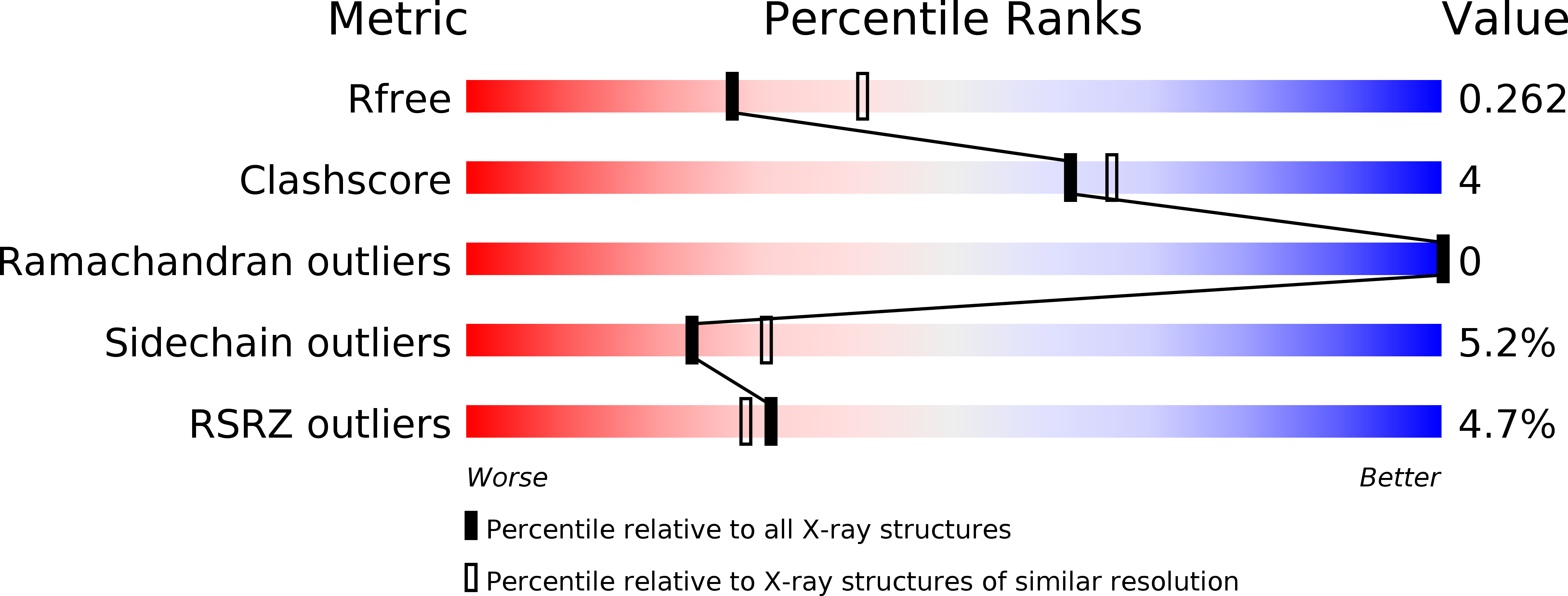
Resolution:
2.44 Å
R-Value Free:
0.26
R-Value Work:
0.19
R-Value Observed:
0.20
Space Group:
C 2 2 21
Macromolecular Entities
Structures with similar UniProt ID
Protein Blast
Polymer Type:polypeptide(L)
Molecule:Synaptobrevin homolog YKT6
Gene (Uniprot):Ykt6
Chain IDs:A
Chain Length:199
Number of Molecules:1
Biological Source:Rattus norvegicus
Primary Citation
Lipid-Induced Conformational Switch Controls Fusion Activity of Longin Domain SNARE Ykt6
Mol.Cell 37 383 395 (2010)
PMID: 20159557 DOI: 10.1016/j.molcel.2010.01.024

Abstact

While most SNAREs are permanently anchored to membranes by their transmembrane domains, the dually lipidated SNARE Ykt6 is found both on intracellular membranes and in the cytosol. The cytosolic Ykt6 is inactive due to the autoinhibition of the SNARE core by its longin domain, although the molecular basis of this inhibition is unknown. Here, we demonstrate that unlipidated Ykt6 adopts multiple conformations, with a small population in the closed state. The structure of Ykt6 in complex with a fatty acid suggests that, upon farnesylation, the Ykt6 SNARE core forms four alpha helices that wrap around the longin domain, forming a dominantly closed conformation. The fatty acid, buried in a hydrophobic groove formed between the longin domain and its SNARE core, is essential for maintaining the autoinhibited conformation of Ykt6. Our study reveals that the posttranslationally attached farnesyl group can actively regulate Ykt6 fusion activity in addition to its anticipated membrane-anchoring role.

Legend

Protein

Chemical

Disease

Primary Citation of related structures