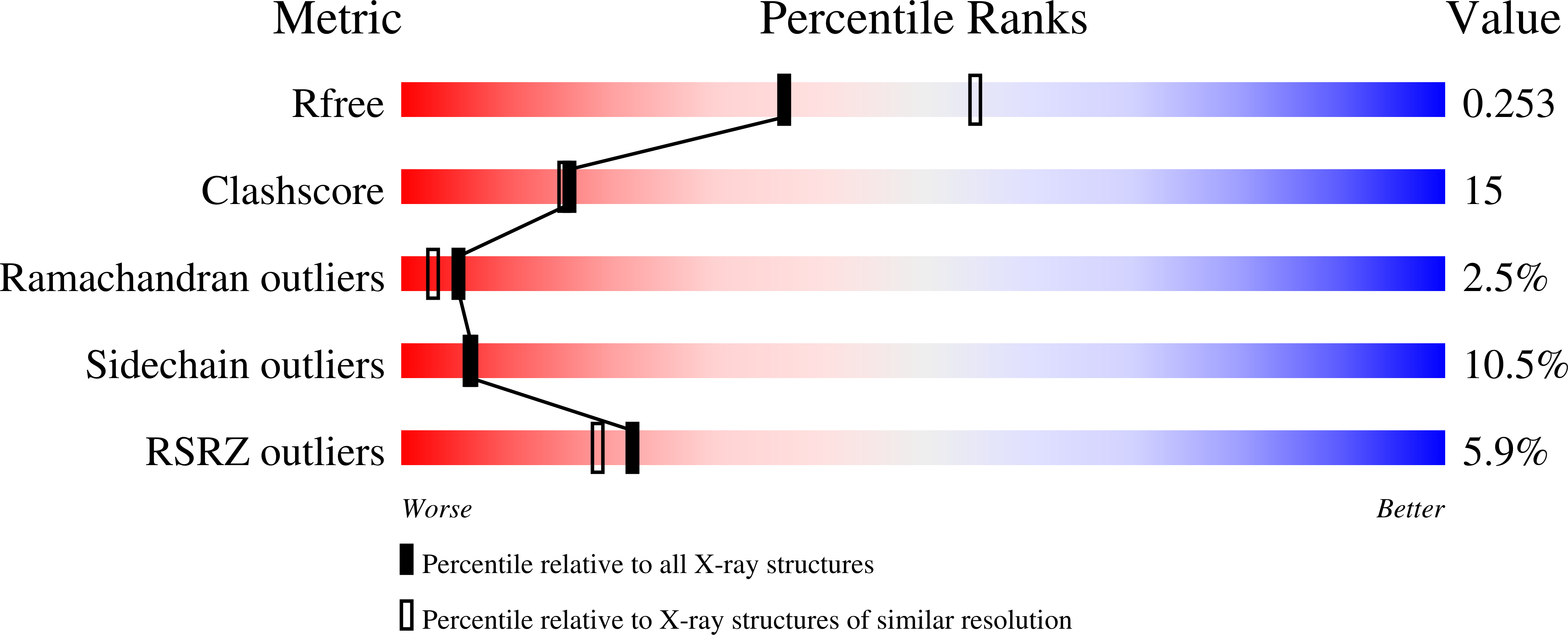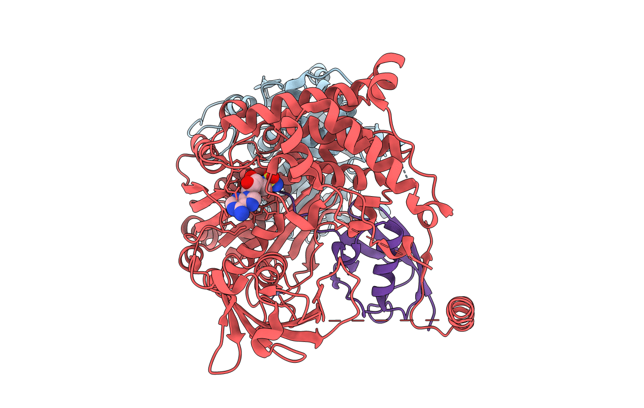
3KYC image
Deposition Date 2009-12-05
Release Date 2010-02-16
Last Version Date 2024-11-06
Entry Detail
PDB ID:
3KYC
Keywords:
Title:
Human SUMO E1 complex with a SUMO1-AMP mimic
Biological Source:
Source Organism:
Homo sapiens (Taxon ID: 9606)
Host Organism:
Method Details:
Experimental Method:
Resolution:
2.45 Å
R-Value Free:
0.25
R-Value Work:
0.19
R-Value Observed:
0.19
Space Group:
P 21 21 21
Macromolecular Entities
Polymer Type:polypeptide(L)
Molecule:SUMO-activating enzyme subunit 1
Gene (Uniprot):SAE1
Chain IDs:A
Chain Length:346
Number of Molecules:1
Biological Source:Homo sapiens
Polymer Type:polypeptide(L)
Molecule:SUMO-activating enzyme subunit 2
Gene (Uniprot):UBA2
Chain IDs:B
Chain Length:660
Number of Molecules:1
Biological Source:Homo sapiens
Polymer Type:polypeptide(L)
Molecule:Small ubiquitin-related modifier 1
Gene (Uniprot):SUMO1
Mutations:T95C
Chain IDs:C (auth: D)
Chain Length:97
Number of Molecules:1
Biological Source:Homo sapiens
Primary Citation
Active site remodelling accompanies thioester bond formation in the SUMO E1.
Nature 463 906 912 (2010)
PMID: 20164921 DOI: 10.1038/nature08765

Abstact

E1 enzymes activate ubiquitin (Ub) and ubiquitin-like (Ubl) proteins in two steps by carboxy-terminal adenylation and thioester bond formation to a conserved catalytic cysteine in the E1 Cys domain. The structural basis for these intermediates remains unknown. Here we report crystal structures for human SUMO E1 in complex with SUMO adenylate and tetrahedral intermediate analogues at 2.45 and 2.6 A, respectively. These structures show that side chain contacts to ATP.Mg are released after adenylation to facilitate a 130 degree rotation of the Cys domain during thioester bond formation that is accompanied by remodelling of key structural elements including the helix that contains the E1 catalytic cysteine, the crossover and re-entry loops, and refolding of two helices that are required for adenylation. These changes displace side chains required for adenylation with side chains required for thioester bond formation. Mutational and biochemical analyses indicate these mechanisms are conserved in other E1s.

Legend

Protein

Chemical

Disease

Primary Citation of related structures