Deposition Date
2009-12-03
Release Date
2010-02-02
Last Version Date
2024-11-06
Entry Detail
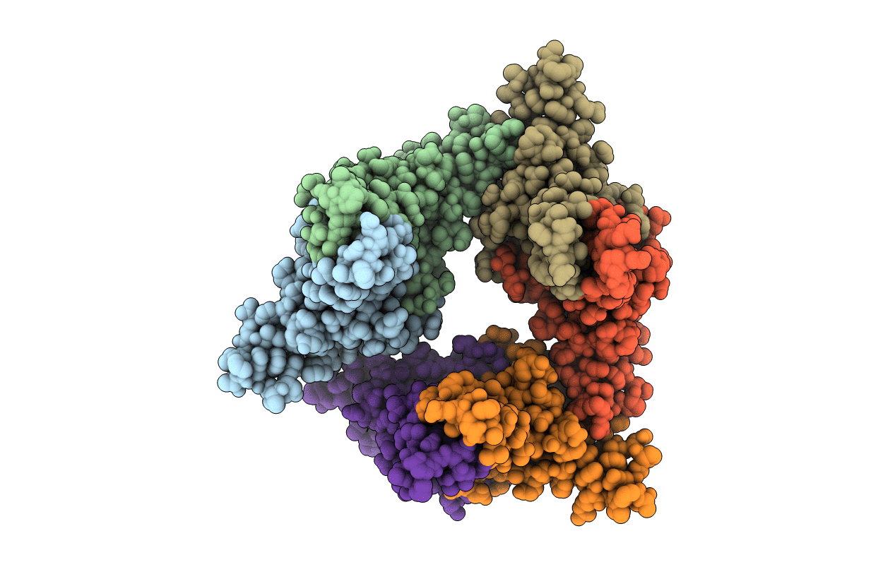
PDB ID:
3KXS
Keywords:
Title:
Crystal structure of HBV capsid mutant dimer (oxy form), strain adyw
Biological Source:
Source Organism(s):
Hepatitis B virus subtype adyw (Taxon ID: 10419)
Expression System(s):
Method Details:
Experimental Method:
Resolution:
2.25 Å
R-Value Free:
0.26
R-Value Work:
0.21
R-Value Observed:
0.21
Space Group:
P 31