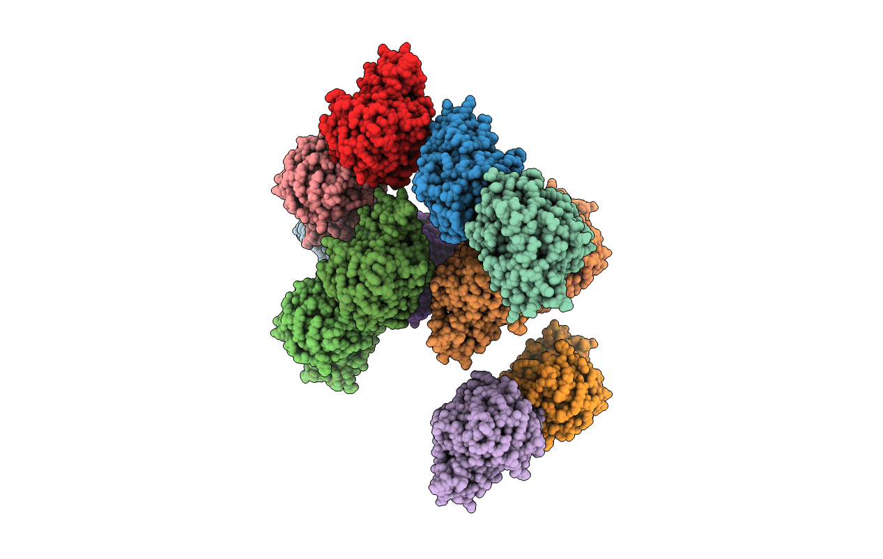
3KXP image
Entry Detail
PDB ID:
3KXP
Keywords:
Title:
Crystal Structure of E-2-(Acetamidomethylene)succinate Hydrolase
Biological Source:
Source Organism:
Host Organism:
PDB Version:
Deposition Date:
2009-12-03
Release Date:
2010-02-09
Method Details:
Experimental Method:
Resolution:
2.26 Å
R-Value Free:
0.24
R-Value Work:
0.20
Space Group:
P 21 21 21
Macromolecular Entities
Polymer Type:polypeptide(L)
Description:Alpha-(N-acetylaminomethylene)succinic acid hydrolase
Chain IDs:A, B, C, D, E, F, G, H, I, J, K, L
Chain Length:314
Number of Molecules:12
Biological Source:Mesorhizobium loti
Modified Residue
Compound ID Chain ID Parent Comp ID Details 2D Image
MSE A MET SELENOMETHIONINE
Ligand Molecules
Primary Citation
Structure determination and characterization of the vitamin B(6) degradative enzyme (E)-2-(acetamidomethylene)succinate hydrolase.
Biochemistry 49 1226 1235 (2010)
PMID: 20099871 DOI: 10.1021/bi901812p

Abstact

The gene identification and kinetic characterization of (E)-2-(acetamidomethylene)succinate (E-2AMS) hydrolase has recently been described. This enzyme catalyzes the final reaction in the degradation of vitamin B(6) and produces succinic semialdehyde, acetate, ammonia, and carbon dioxide from E-2AMS. The structure of E-2AMS hydrolase was determined to 2.3 A using SAD phasing. E-2AMS hydrolase is a member of the alpha/beta hydrolase superfamily and utilizes a serine/histidine/aspartic acid catalytic triad. Mutation of either the nucleophilic serine or the aspartate resulted in inactive enzyme. Mutation of an additional serine residue in the active site causes the enzyme to be unstable and is likely structurally important. The structure also provides insight into the mechanism of hydrolysis of E-2AMS and identifies several potential catalytically important residues.

Legend

Protein

Chemical

Disease

Primary Citation of related structures