3KX9 image
Deposition Date 2009-12-02
Release Date 2010-12-15
Last Version Date 2023-09-06
Entry Detail
PDB ID:
3KX9
Title:
Engineering a closed form of the Archaeoglobus fulgidus ferritin by site directed mutagenesis
Biological Source:
Source Organism:
Host Organism:
Method Details:
Experimental Method:
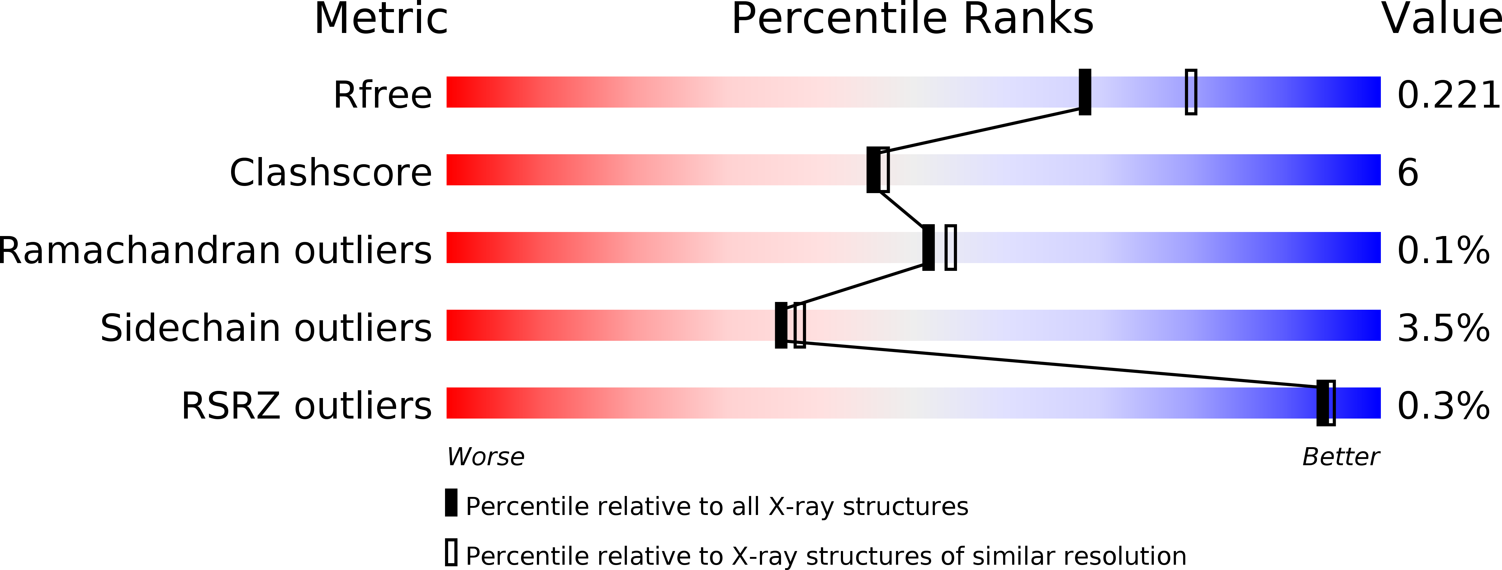
Resolution:
2.10 Å
R-Value Free:
0.22
R-Value Work:
0.18
R-Value Observed:
0.18
Space Group:
P 21 21 21
Macromolecular Entities
Structures with similar UniProt ID
Protein Blast
Polymer Type:polypeptide(L)
Molecule:Ferritin
Gene (Uniprot):AF_0834
Mutagens:K150A, R151A
Chain IDs:A, B, C, D, E, F, G, H, I, J, K, L, M, N, O, P, Q, R, S, T, U, V, W, X
Chain Length:173
Number of Molecules:24
Biological Source:Archaeoglobus fulgidus
Ligand Molecules
Primary Citation
The role of nonconserved residues of Archaeoglobus fulgidus ferritin on its unique structure and biophysical properties.
J.Biol.Chem. 288 32663 32672 (2013)
PMID: 24030827 DOI: 10.1074/jbc.M113.491191

Abstact

Archaeoglobus fulgidus ferritin (AfFtn) is the only tetracosameric ferritin known to form a tetrahedral cage, a structure that remains unique in structural biology. As a result of the tetrahedral (2-3) symmetry, four openings (∼45 Å in diameter) are formed in the cage. This open tetrahedral assembly contradicts the paradigm of a typical ferritin cage: a closed assembly having octahedral (4-3-2) symmetry. To investigate the molecular mechanism affecting this atypical assembly, amino acid residues Lys-150 and Arg-151 were replaced by alanine. The data presented here shed light on the role that these residues play in shaping the unique structural features and biophysical properties of the AfFtn. The x-ray crystal structure of the K150A/R151A mutant, solved at 2.1 Å resolution, indicates that replacement of these key residues flips a "symmetry switch." The engineered molecule no longer assembles with tetrahedral symmetry but forms a typical closed octahedral ferritin cage. Small angle x-ray scattering reveals that the overall shape and size of AfFtn and AfFtn-AA in solution are consistent with those observed in their respective crystal structures. Iron binding and release kinetics of the AfFtn and AfFtn-AA were investigated to assess the contribution of cage openings to the kinetics of iron oxidation, mineralization, or reductive iron release. Identical iron binding kinetics for AfFtn and AfFtn-AA suggest that Fe(2+) ions do not utilize the triangular pores for access to the catalytic site. In contrast, relatively slow reductive iron release was observed for the closed AfFtn-AA, demonstrating involvement of the large pores in the pathway for iron release.

Legend

Protein

Chemical

Disease

Primary Citation of related structures