3KUT image
Deposition Date 2009-11-27
Release Date 2010-02-09
Last Version Date 2023-09-06
Entry Detail
PDB ID:
3KUT
Keywords:
Title:
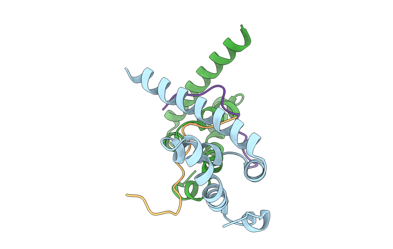
Crystal structure of the MLLE domain of poly(A)-binding protein in complex with the binding region of Paip2
Biological Source:
Source Organism:
Homo sapiens (Taxon ID: 9606)
Host Organism:
Method Details:
Experimental Method:
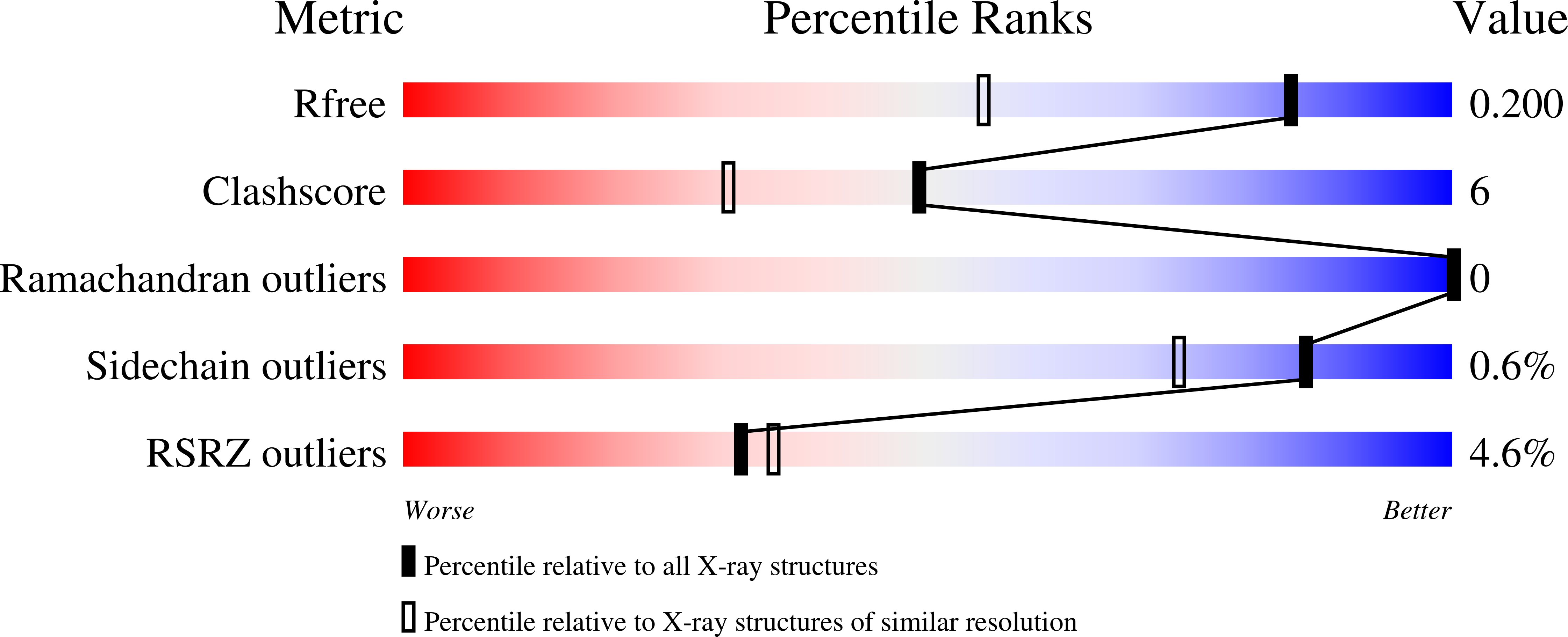
Resolution:
1.50 Å
R-Value Free:
0.20
R-Value Work:
0.16
R-Value Observed:
0.16
Space Group:
P 1
Macromolecular Entities
Polymer Type:polypeptide(L)
Molecule:Polyadenylate-binding protein 1
Gene (Uniprot):PABPC1
Chain IDs:A, B
Chain Length:88
Number of Molecules:2
Biological Source:Homo sapiens
Polymer Type:polypeptide(L)
Molecule:PAIP2 protein
Mutations:K116A
Chain IDs:C, D
Chain Length:17
Number of Molecules:2
Biological Source:Homo sapiens
Ligand Molecules
Primary Citation
Molecular Determinants of PAM2 Recognition by the MLLE Domain of Poly(A)-Binding Protein.
J.Mol.Biol. 397 397 407 (2010)
PMID: 20096703 DOI: 10.1016/j.jmb.2010.01.032

Abstact

MLLE (previously known as PABC) is a peptide-binding domain that is found in poly(A)-binding protein (PABP) and EDD (E3 isolated by differential display), a HECT E3 ubiquitin ligase also known as HYD (hyperplastic discs tumor suppressor) or UBR5. The MLLE domain from PABP recruits various regulatory proteins and translation factors to poly(A) mRNAs through binding of a conserved 12 amino acid peptide motif called PAM2 (for PABP-interacting motif 2). Here, we determined crystal structures of the MLLE domain from PABP alone and in complex with PAM2 peptides from PABP-interacting protein 2. The structures provide a detailed view of hydrophobic determinants of the MLLE binding coded by PAM2 positions 3, 5, 7, 10, and 12 and reveal novel intermolecular polar contacts. In particular, the side chain of the invariant MLLE residue K580 forms hydrogen bonds with the backbone of PAM2 residues 5 and 7. The structures also show that peptide residues outside of the conserved PAM2 motif contribute to binding. Altogether, the structures provide a significant advance in understanding the molecular basis for the binding of PABP by PAM2-containing proteins involved in translational control, mRNA deadenylation, and other cellular processes.

Legend

Protein

Chemical

Disease

Primary Citation of related structures