Deposition Date
2009-11-20
Release Date
2010-05-26
Last Version Date
2024-11-20
Entry Detail
PDB ID:
3KS0
Keywords:
Title:
Crystal structure of the heme domain of flavocytochrome b2 in complex with Fab B2B4
Biological Source:
Source Organism(s):
Saccharomyces cerevisiae (Taxon ID: 4932)
Mus musculus (Taxon ID: 10090)
Mus musculus (Taxon ID: 10090)
Expression System(s):
Method Details:
Experimental Method:
Resolution:
2.70 Å
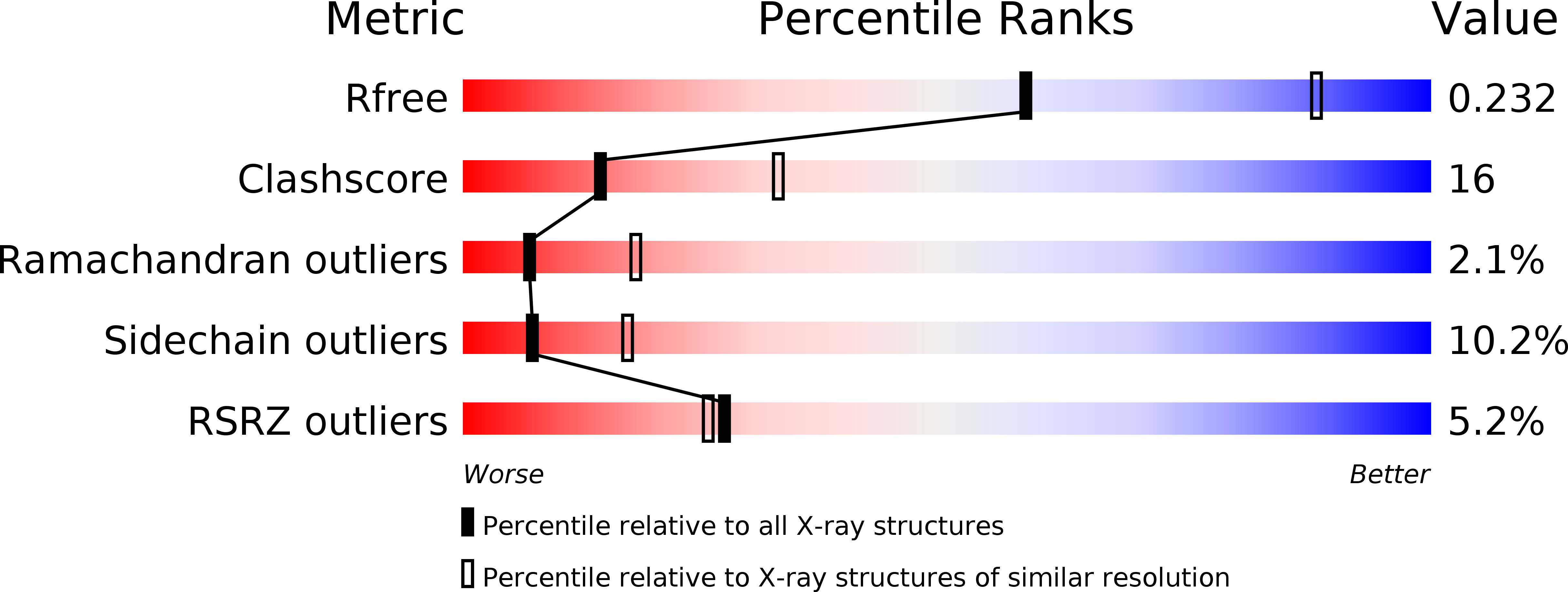
R-Value Free:
0.28
R-Value Work:
0.21
R-Value Observed:
0.21
Space Group:
P 1 21 1