3KP9 image
Deposition Date 2009-11-16
Release Date 2010-02-09
Last Version Date 2024-11-20
Entry Detail
PDB ID:
3KP9
Title:
Structure of a bacterial homolog of vitamin K epoxide reductase
Biological Source:
Source Organism:
Host Organism:
Method Details:
Experimental Method:
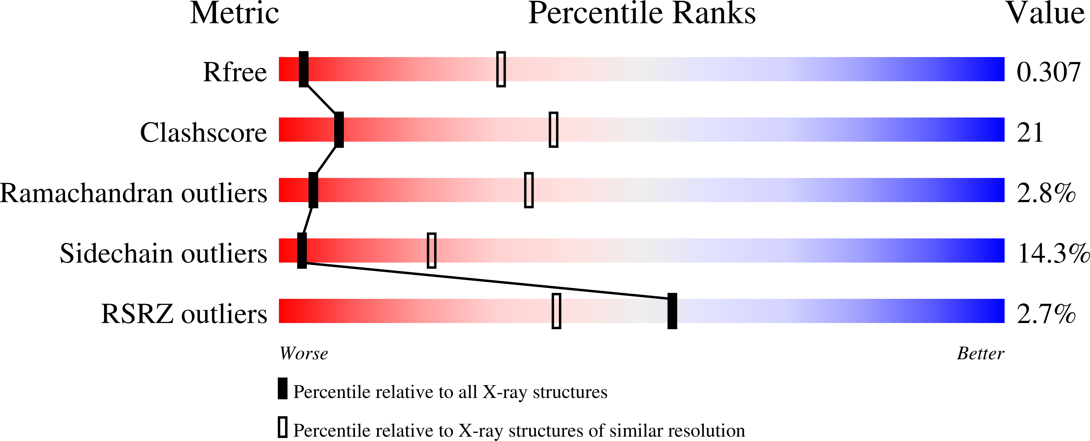
Resolution:
3.60 Å
R-Value Free:
0.30
R-Value Work:
0.25
R-Value Observed:
0.25
Space Group:
P 61
Macromolecular Entities
Structures with similar UniProt ID
Protein Blast
Polymer Type:polypeptide(L)
Molecule:VKORC1/thioredoxin domain protein
Gene (Uniprot):CYB_2278
Mutagens:C56S
Chain IDs:A
Chain Length:291
Number of Molecules:1
Biological Source:Synechococcus sp.
Primary Citation
Structure of a bacterial homologue of vitamin K epoxide reductase.
Nature 463 507 512 (2010)
PMID: 20110994 DOI: 10.1038/nature08720

Abstact

Vitamin K epoxide reductase (VKOR) generates vitamin K hydroquinone to sustain gamma-carboxylation of many blood coagulation factors. Here, we report the 3.6 A crystal structure of a bacterial homologue of VKOR from Synechococcus sp. The structure shows VKOR in complex with its naturally fused redox partner, a thioredoxin-like domain, and corresponds to an arrested state of electron transfer. The catalytic core of VKOR is a four transmembrane helix bundle that surrounds a quinone, connected through an additional transmembrane segment with the periplasmic thioredoxin-like domain. We propose a pathway for how VKOR uses electrons from cysteines of newly synthesized proteins to reduce a quinone, a mechanism confirmed by in vitro reconstitution of vitamin K-dependent disulphide bridge formation. Our results have implications for the mechanism of the mammalian VKOR and explain how mutations can cause resistance to the VKOR inhibitor warfarin, the most commonly used oral anticoagulant.

Legend

Protein

Chemical

Disease

Primary Citation of related structures
Feedback Form
Name
Email
Institute
Feedback