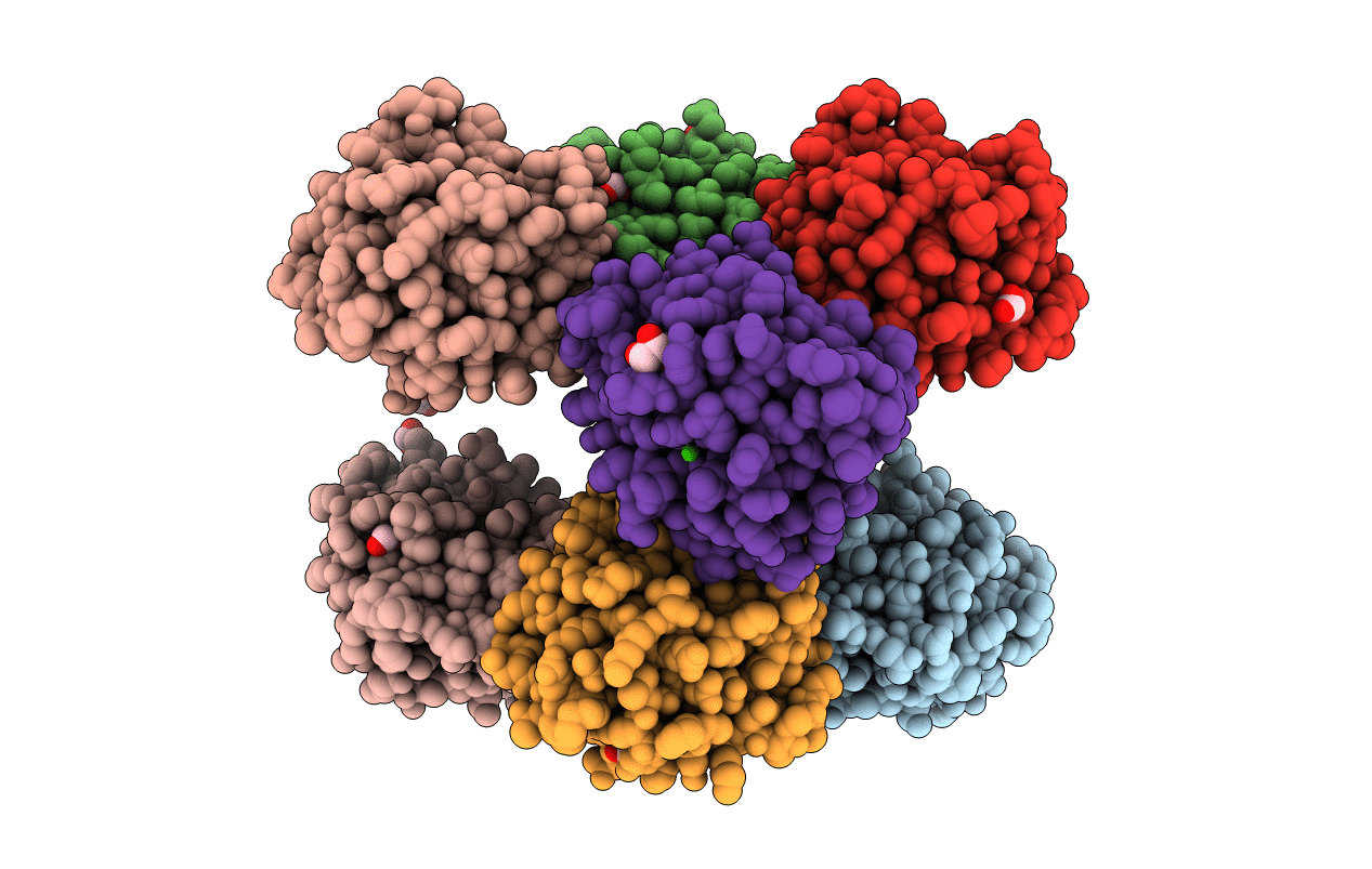
3KMV image
Deposition Date 2009-11-11
Release Date 2010-08-04
Last Version Date 2023-09-06
Entry Detail
PDB ID:
3KMV
Title:
Crystal structure of CBM42A from Clostridium thermocellum
Biological Source:
Source Organism:
Method Details:
Experimental Method:
Resolution:
1.80 Å
R-Value Free:
0.19
R-Value Work:
0.15
R-Value Observed:
0.16
Space Group:
P 32 2 1
Macromolecular Entities
Structures with similar UniProt ID
Protein Blast
Polymer Type:polypeptide(L)
Molecule:Alpha-L-arabinofuranosidase B
Gene (Uniprot):Cthe_0015
Chain IDs:A, B, C, D, E, F, G, H
Chain Length:157
Number of Molecules:8
Biological Source:Clostridium thermocellum
Primary Citation
Family 42 carbohydrate-binding modules display multiple arabinoxylan-binding interfaces presenting different ligand affinities.
Biochim.Biophys.Acta 1804 2054 2062 (2010)
PMID: 20637315 DOI: 10.1016/j.bbapap.2010.07.006

Abstact

Enzymes that degrade plant cell wall polysaccharides display a modular architecture comprising a catalytic domain bound to one or more non-catalytic carbohydrate-binding modules (CBMs). CBMs display considerable variation in primary structure and are grouped into 59 sequence-based families organized in the Carbohydrate-Active enZYme (CAZy) database. Here we report the crystal structure of CtCBM42A together with the biochemical characterization of two other members of family 42 CBMs from Clostridium thermocellum. CtCBM42A, CtCBM42B and CtCBM42C bind specifically to the arabinose side-chains of arabinoxylans and arabinan, suggesting that various cellulosomal components are targeted to these regions of the plant cell wall. The structure of CtCBM42A displays a beta-trefoil fold, which comprises 3 sub-domains designated as alpha, beta and gamma. Each one of the three sub-domains presents a putative carbohydrate-binding pocket where an aspartate residue located in a central position dominates ligand recognition. Intriguingly, the gamma sub-domain of CtCBM42A is pivotal for arabinoxylan binding, while the concerted action of beta and gamma sub-domains of CtCBM42B and CtCBM42C is apparently required for ligand sequestration. Thus, this work reveals that the binding mechanism of CBM42 members is in contrast with that of homologous CBM13s where recognition of complex polysaccharides results from the cooperative action of three protein sub-domains presenting similar affinities.

Legend

Protein

Chemical

Disease

Primary Citation of related structures