Deposition Date
2009-11-10
Release Date
2010-03-23
Last Version Date
2023-11-01
Entry Detail
PDB ID:
3KM6
Keywords:
Title:
Crystal Structure of the Human GST Pi C47S/Y108V Double Mutant in Complex with the Ethacrynic Acid-Glutathione Conjugate
Biological Source:
Source Organism(s):
Homo sapiens (Taxon ID: 9606)
Expression System(s):
Method Details:
Experimental Method:
Resolution:
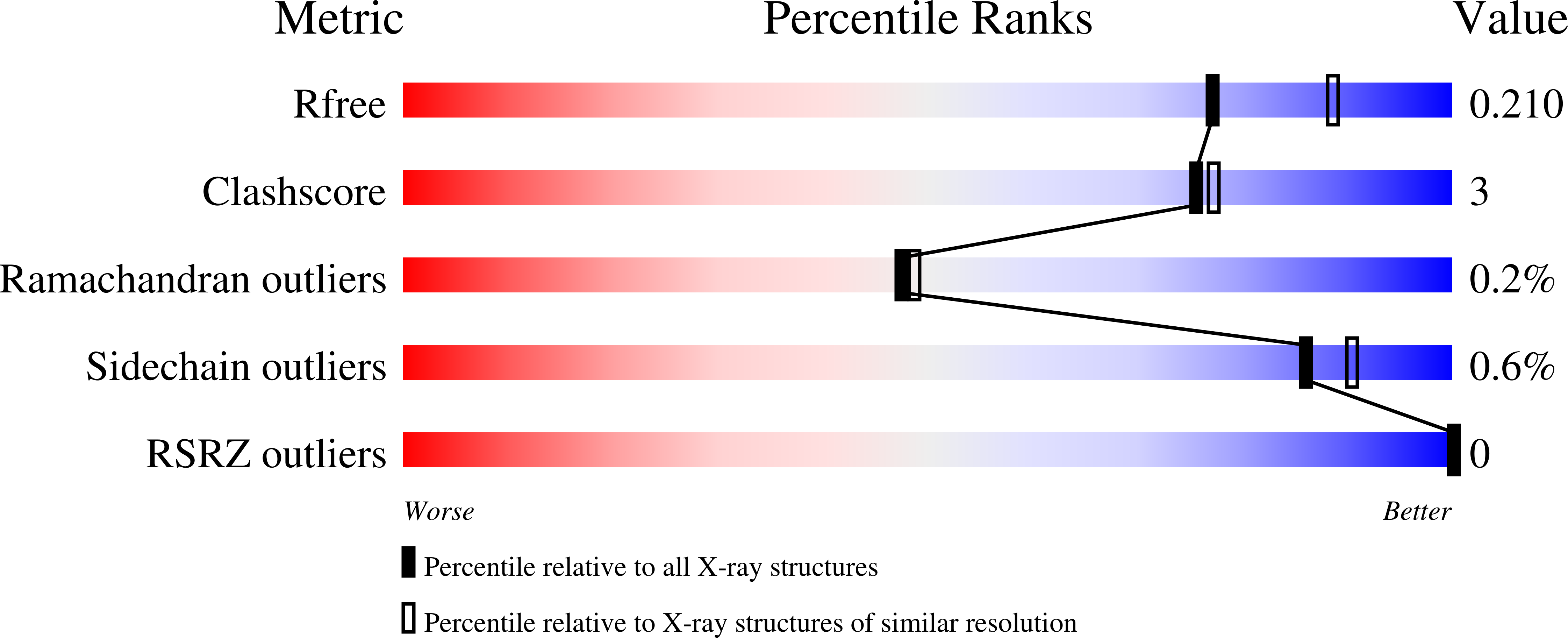
2.10 Å
R-Value Free:
0.20
R-Value Work:
0.15
R-Value Observed:
0.15
Space Group:
C 1 2 1