3KJR image
Deposition Date 2009-11-03
Release Date 2009-11-17
Last Version Date 2023-09-06
Entry Detail
PDB ID:
3KJR
Keywords:
Title:
Crystal structure of dihydrofolate reductase/thymidylate synthase from Babesia bovis determined using SlipChip based microfluidics
Biological Source:
Source Organism:
Method Details:
Experimental Method:
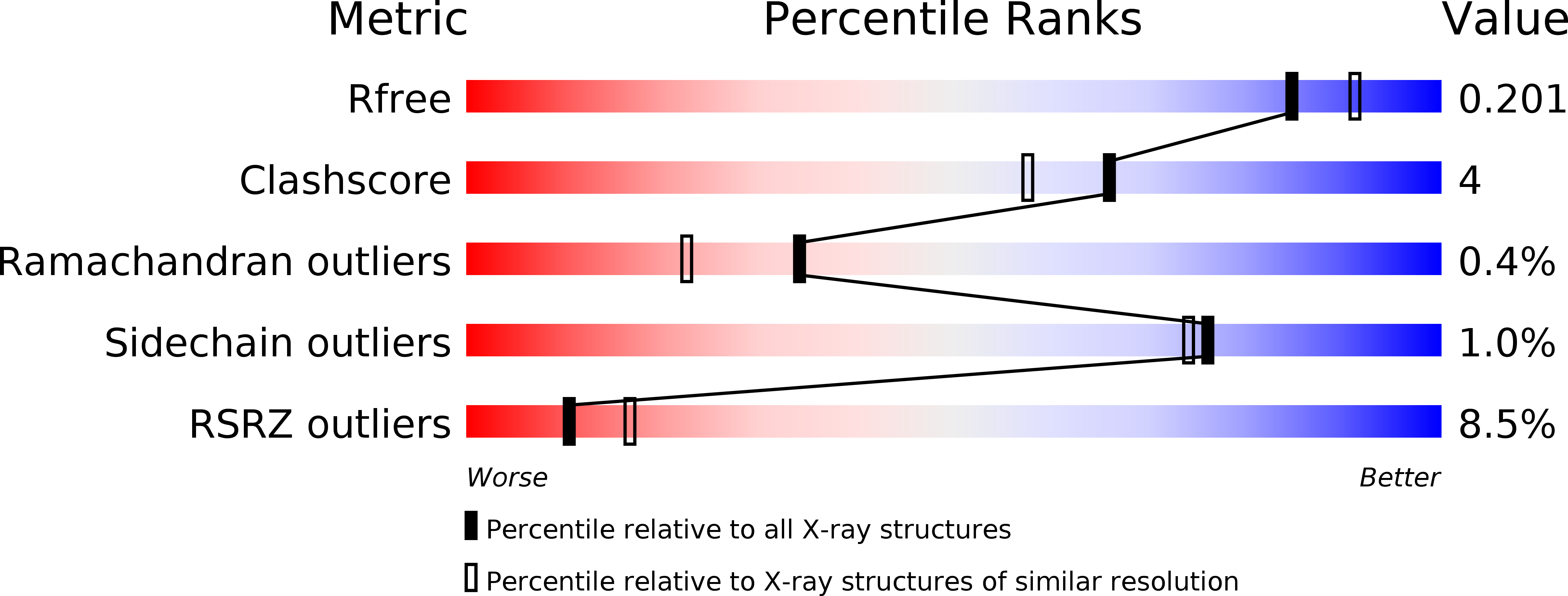
Resolution:
1.95 Å
R-Value Free:
0.20
R-Value Work:
0.16
R-Value Observed:
0.17
Space Group:
P 21 21 21
Macromolecular Entities
Structures with similar UniProt ID
Protein Blast
Polymer Type:polypeptide(L)
Molecule:Dihydrofolate reductase/thymidylate synthase
Gene (Uniprot):BBOV_II000780
Chain IDs:A, B
Chain Length:511
Number of Molecules:2
Biological Source:Babesia bovis T2Bo
Primary Citation
Multiparameter screening on SlipChip used for nanoliter protein crystallization combining free interface diffusion and microbatch methods.
J.Am.Chem.Soc. 132 112 119 (2010)
PMID: 20000709 DOI: 10.1021/ja908558m

Abstact

This paper describes two SlipChip-based approaches to protein crystallization: a SlipChip-based free interface diffusion (FID) method and a SlipChip-based composite method that simultaneously performs microbatch and FID crystallization methods in a single device. The FID SlipChip was designed to screen multiple reagents, each at multiple diffusion equilibration times, and was validated by screening conditions for crystallization of two proteins, enoyl-CoA hydratase from Mycobacterium tuberculosis and dihydrofolate reductase/thymidylate synthase from Babesia bovis, against 48 different reagents at five different equilibration times each, consuming 12 microL of each protein for a total of 480 experiments using three SlipChips. The composite SlipChip was designed to screen multiple reagents, each at multiple mixing ratios and multiple equilibration times, and was validated by screening conditions for crystallization of two proteins, enoyl-CoA hydratase from Mycobacterium tuberculosis and dihydrofolate reductase/thymidylate synthase from Babesia bovis. To prevent cross-contamination while keeping the solution in the neck channels for FID stable, the plates of the SlipChip were etched with a pattern of nanowells. This nanopattern was used to increase the contact angle of aqueous solutions on the surface of the silanized glass. The composite SlipChip increased the number of successful crystallization conditions and identified more conditions for crystallization than separate FID and microbatch screenings. Crystallization experiments were scaled up in well plates using conditions identified during the SlipChip screenings, and X-ray diffraction data were obtained to yield the protein structure of dihydrofolate reductase/thymidylate synthase at 1.95 A resolution. This free-interface diffusion approach provides a convenient and high-throughput method of setting up gradients in microfluidic devices and may find additional applications in cell-based assays.

Legend

Protein

Chemical

Disease

Primary Citation of related structures
Feedback Form
Name
Email
Institute
Feedback