3KJ7 image
Deposition Date 2009-11-03
Release Date 2010-02-16
Last Version Date 2024-11-20
Entry Detail
PDB ID:
3KJ7
Keywords:
Title:
Crystal Structure of the Complex of C-lobe of Bovine Lactoferrin with Dextrin at 1.9 A Resolution
Biological Source:
Source Organism:
Bos taurus (Taxon ID: 9913)
Method Details:
Experimental Method:
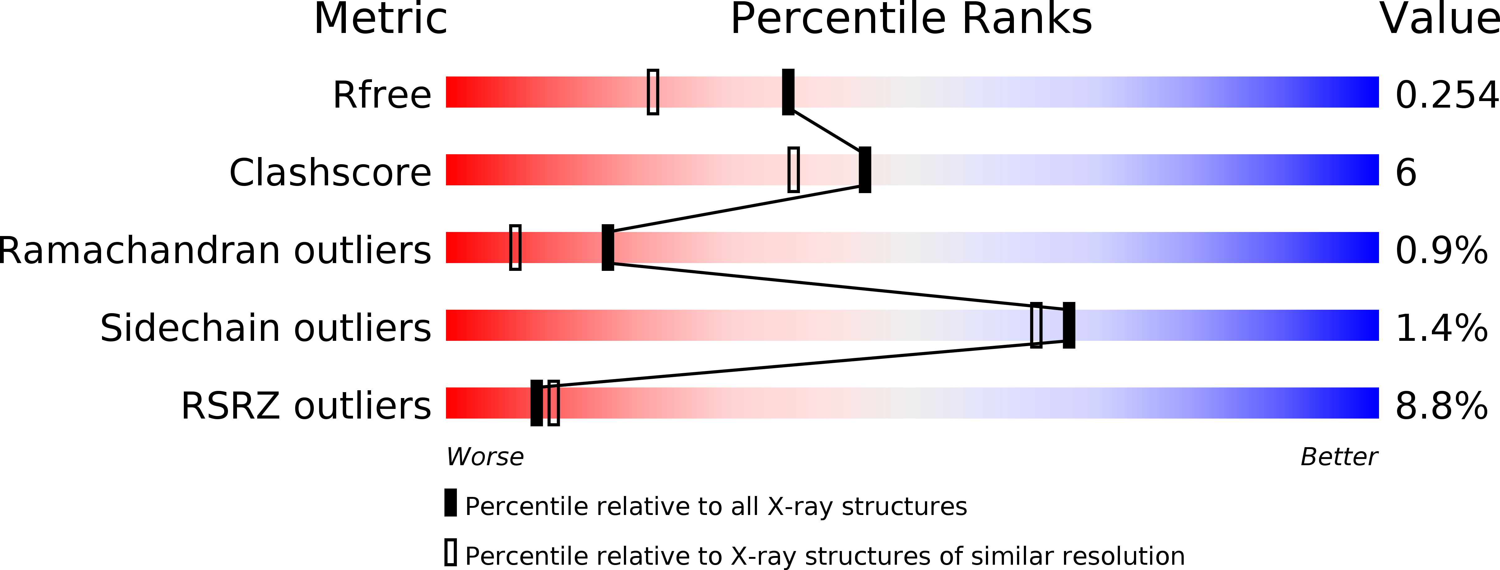
Resolution:
1.91 Å
R-Value Free:
0.24
R-Value Work:
0.20
R-Value Observed:
0.21
Space Group:
P 1 21 1
Macromolecular Entities
Structures with similar UniProt ID
Protein Blast
Polymer Type:polypeptide(L)
Molecule:Lactotransferrin
Gene (Uniprot):LTF
Chain IDs:A
Chain Length:345
Number of Molecules:1
Biological Source:Bos taurus
Modified Residue
Compound ID Chain ID Parent Comp ID Details 2D Image
ASN A ASN GLYCOSYLATION SITE
Peptide-like Molecules
PRD_900014
Primary Citation
Specific interactions of C-terminal half (C-lobe) of lactoferrin protein with edible sugars: binding and structural studies with implications on diabetes.
Int.J.Biol.Macromol. 47 50 59 (2010)
PMID: 20371371 DOI: 10.1016/j.ijbiomac.2010.03.021

Abstact

Bovine lactoferrin has been shown to reduce the levels of glucose in both normal subjects and non-insulin dependent diabetic patients. The binding studies have shown that various sugar molecules interact with lactoferrin indicating the presence of a sugar-binding site in the protein. Structural studies have revealed that the sugar-binding site is located in the C-terminal half (C-lobe) of bilobal lactoferrin. Since the sugar-binding site was part of the C-lobe, it was better to carry out binding and structural studies using C-lobe rather than the full protein molecule. Therefore, C-lobe was prepared by limited proteolysis of lactoferrin with enzyme proteinase K. It was purified to homogeneity for further studies. The addition of C-lobe to human serum showed significant lowering of glucose levels. The binding studies using C-lobe with nine sugars, glucose, galactose, mannose, xylose, maltose, cellobiose, lactose, sucrose and dextrin gave values of binding constants in the range of 10(-4) to 10(-5)M. The structure determinations of the complexes of C-lobe with all the nine sugars showed that all of them interact with C-lobe through the same recognition site involving several hydrogen bonds and van der Waals interactions.

Legend

Protein

Chemical

Disease

Primary Citation of related structures