Deposition Date
2009-11-02
Release Date
2010-02-16
Last Version Date
2024-11-06
Entry Detail
PDB ID:
3KJ6
Keywords:
Title:
Crystal structure of a Methylated beta2 Adrenergic Receptor-Fab complex
Biological Source:
Source Organism(s):
Homo sapiens (Taxon ID: 9606)
Mus musculus (Taxon ID: 10090)
Mus musculus (Taxon ID: 10090)
Expression System(s):
Method Details:
Experimental Method:
Resolution:
3.40 Å
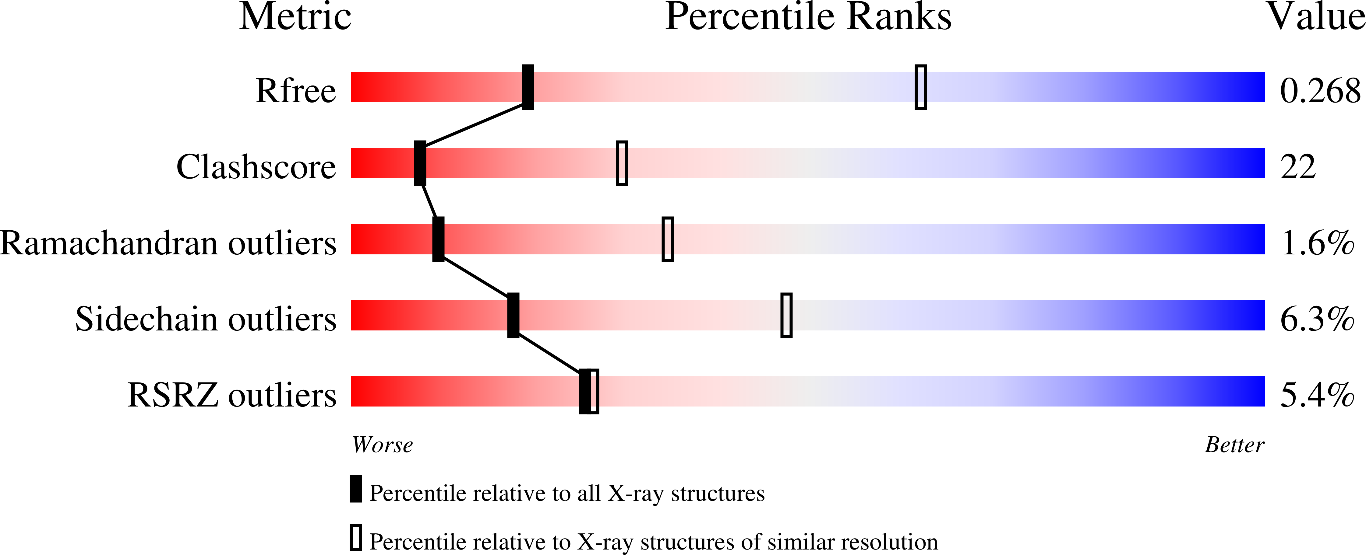
R-Value Free:
0.28
R-Value Work:
0.23
R-Value Observed:
0.24
Space Group:
C 1 2 1