3KG7 image
Deposition Date 2009-10-28
Release Date 2010-01-19
Last Version Date 2024-10-09
Entry Detail
PDB ID:
3KG7
Keywords:
Title:
Dehydratase domain from CurH module of Curacin polyketide synthase
Biological Source:
Source Organism:
Host Organism:
Method Details:
Experimental Method:
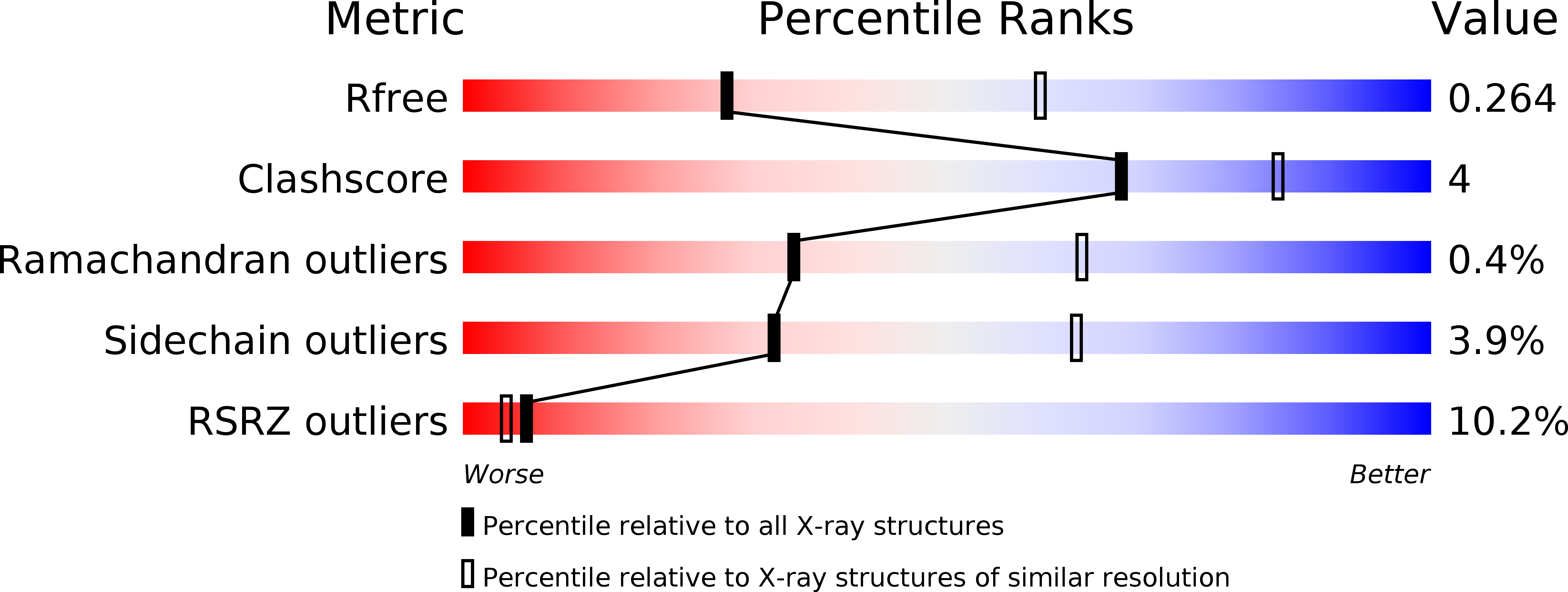
Resolution:
2.77 Å
R-Value Free:
0.26
R-Value Work:
0.20
R-Value Observed:
0.20
Space Group:
P 1 21 1
Macromolecular Entities
Polymer Type:polypeptide(L)
Molecule:CurH
Gene (Uniprot):curH
Chain IDs:A, B, C, D
Chain Length:293
Number of Molecules:4
Biological Source:Lyngbya majuscula
Modified Residue
Compound ID Chain ID Parent Comp ID Details 2D Image
MSE A MET SELENOMETHIONINE
Primary Citation
Crystal Structures of Dehydratase Domains from the Curacin Polyketide Biosynthetic Pathway.
Structure 18 94 105 (2010)
PMID: 20152156 DOI: 10.1016/j.str.2009.10.018

Abstact

Modular polyketide synthases (PKS) make novel natural products through a series of preprogrammed chemical steps catalyzed by an assembly line of multidomain modules. Each assembly-line step involves unique extension and modification reactions, resulting in tremendous diversity of polyketide products. Dehydratase domains catalyze formation of an alpha,beta-double bond in the nascent polyketide intermediate. We present crystal structures of the four dehydratase domains from the curacin A PKS. The catalytic residues and substrate binding site reside in a tunnel within a single monomer. The positions of the catalytic residues and shape of the substrate tunnel explain how chirality of the substrate hydroxyl group may determine the configuration of the product double bond. Access to the active site may require opening the substrate tunnel, forming an open trench. The arrangement of monomers within the dimer is consistent among PKS dehydratases and differs from that seen in the related mammalian fatty acid synthases.

Legend

Protein

Chemical

Disease

Primary Citation of related structures