Deposition Date
2009-10-27
Release Date
2010-06-23
Last Version Date
2024-10-30
Entry Detail
PDB ID:
3KFE
Keywords:
Title:
Crystal structures of a group II chaperonin from Methanococcus maripaludis
Biological Source:
Source Organism(s):
Methanococcus maripaludis (Taxon ID: 39152)
Expression System(s):
Method Details:
Experimental Method:
Resolution:
3.50 Å
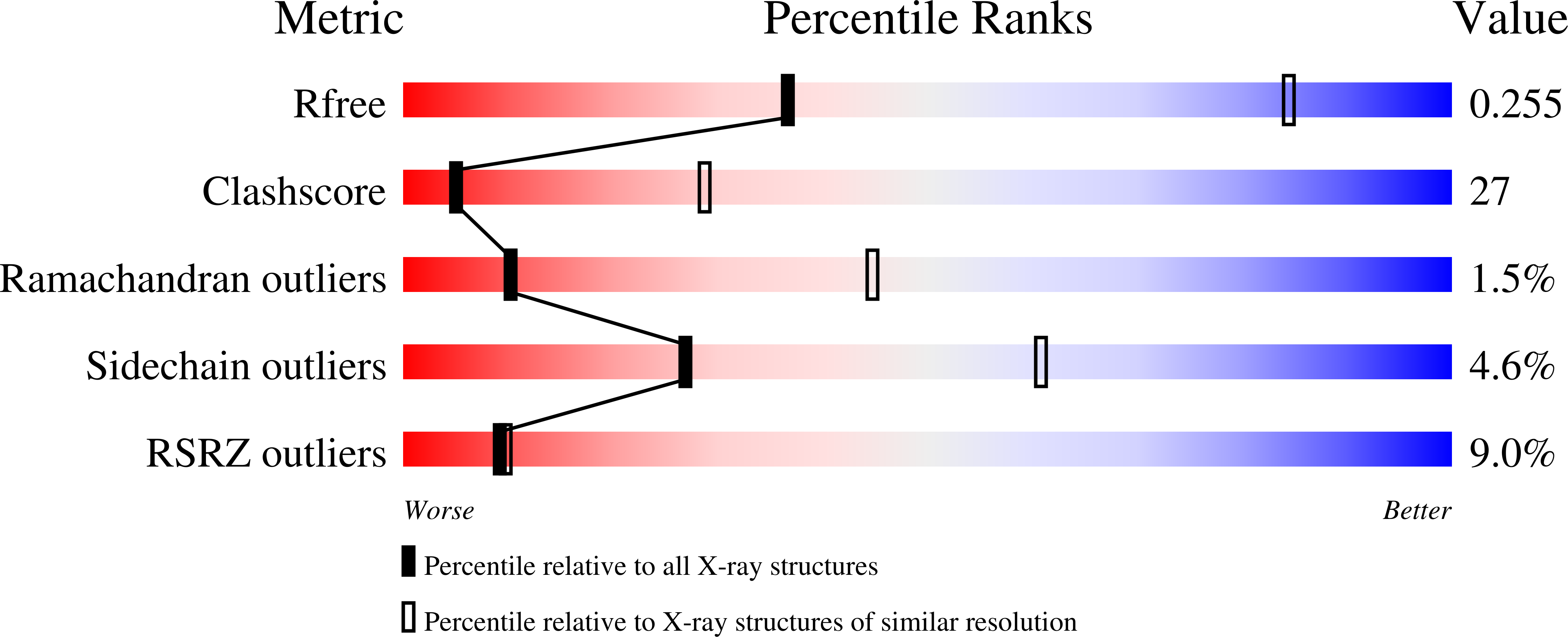
R-Value Free:
0.26
R-Value Work:
0.23
R-Value Observed:
0.23
Space Group:
C 1 2 1