Deposition Date
2009-10-23
Release Date
2009-12-01
Last Version Date
2023-11-01
Entry Detail
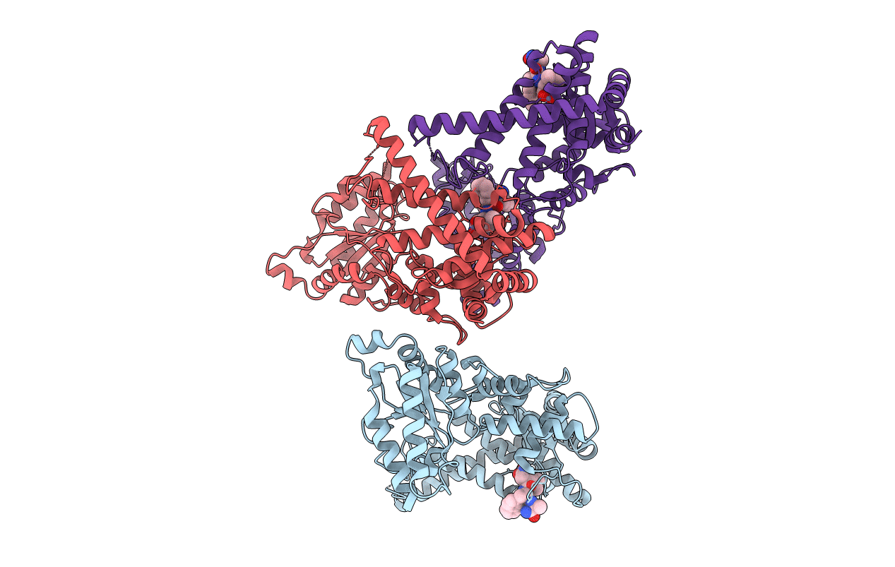
Biological Source:
Source Organism:
Thermotoga maritima (Taxon ID: 2336)
Host Organism:
Method Details:
Experimental Method:
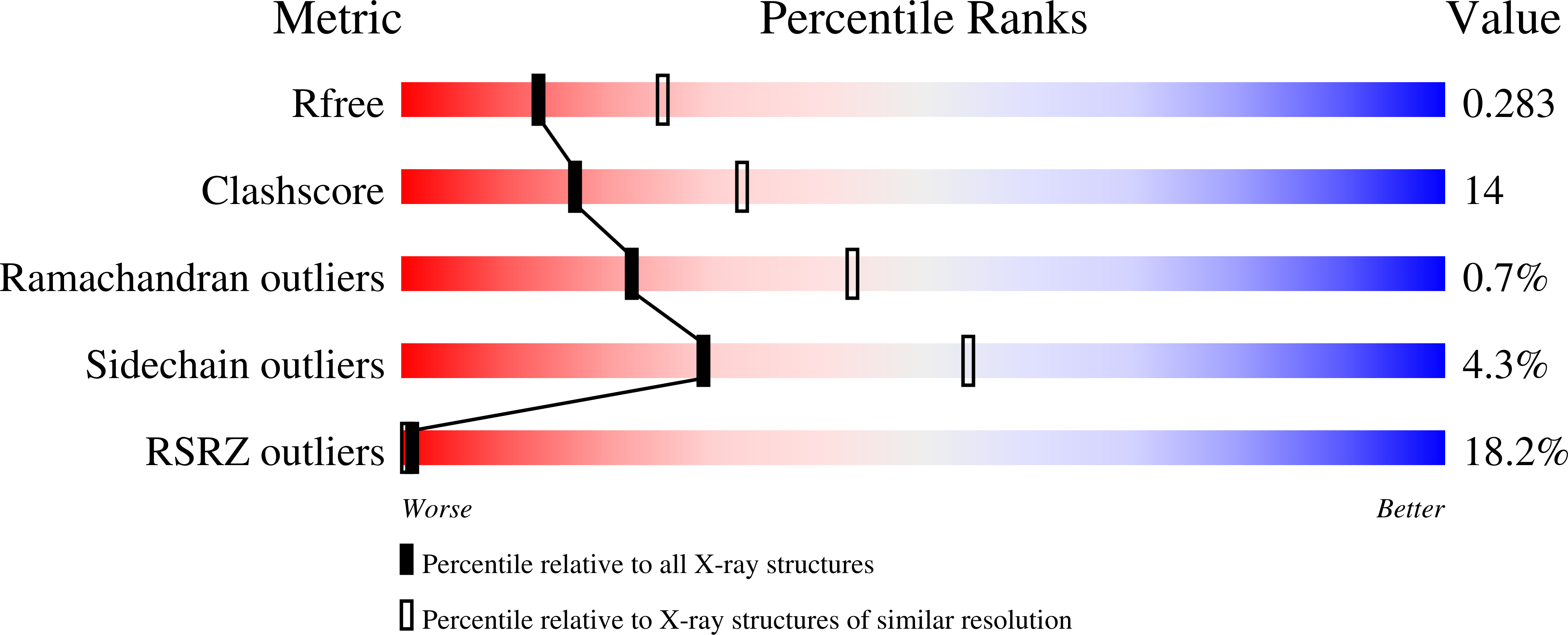
Resolution:
2.60 Å
R-Value Free:
0.28
R-Value Work:
0.22
R-Value Observed:
0.22
Space Group:
P 6 2 2