3KDO image
Entry Detail
PDB ID:
3KDO
Keywords:
Title:
Crystal structure of Type III Rubisco SP6 mutant complexed with 2-CABP
Biological Source:
Host Organism:
PDB Version:
Deposition Date:
2009-10-23
Release Date:
2010-10-06
Method Details:
Experimental Method:
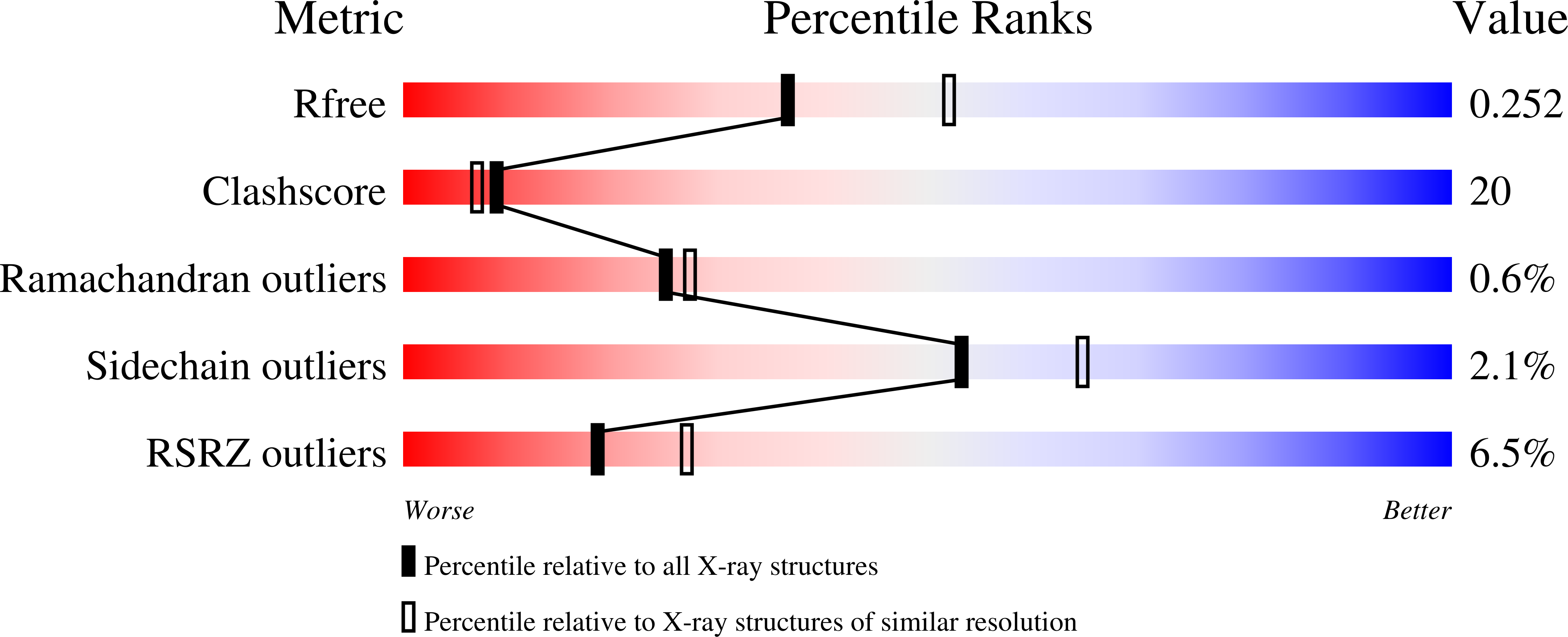
Resolution:
2.36 Å
R-Value Free:
0.26
R-Value Work:
0.21
R-Value Observed:
0.22
Space Group:
P 1 21 1
Macromolecular Entities
Polymer Type:polypeptide(L)
Description:Ribulose bisphosphate carboxylase
Mutations:G326E, K327R, W328D, D329I, V330T, I331L, Q332G, N333F, A334V, R335D, I336L
Chain IDs:A, B, C, D, E, F, G, H, I, J
Chain Length:444
Number of Molecules:10
Biological Source:Thermococcus kodakaraensis
Modified Residue
Compound ID Chain ID Parent Comp ID Details 2D Image
KCX A LYS LYSINE NZ-CARBOXYLIC ACID
Primary Citation
Structure-based catalytic optimization of a type III Rubisco from a hyperthermophile
J.Biol.Chem. 285 39339 39347 (2010)
PMID: 20926376 DOI: 10.1074/jbc.M110.147587

Abstact

The Calvin-Benson-Bassham cycle is responsible for carbon dioxide fixation in all plants, algae, and cyanobacteria. The enzyme that catalyzes the carbon dioxide-fixing reaction is ribulose-1,5-bisphosphate carboxylase/oxygenase (Rubisco). Rubisco from a hyperthermophilic archaeon Thermococcus kodakarensis (Tk-Rubisco) belongs to the type III group, and shows high activity at high temperatures. We have previously found that replacement of the entire α-helix 6 of Tk-Rubisco with the corresponding region of the spinach enzyme (SP6 mutant) results in an improvement of catalytic performance at mesophilic temperatures, both in vivo and in vitro, whereas the former and latter half-replacements of the α-helix 6 (SP4 and SP5 mutants) do not yield such improvement. We report here the crystal structures of the wild-type Tk-Rubisco and the mutants SP4 and SP6, and discuss the relationships between their structures and enzymatic activities. A comparison among these structures shows the movement and the increase of temperature factors of α-helix 6 induced by four essential factors. We thus supposed that an increase in the flexibility of the α-helix 6 and loop 6 regions was important to increase the catalytic activity of Tk-Rubisco at ambient temperatures. Based on this structural information, we constructed a new mutant, SP5-V330T, which was designed to have significantly greater flexibility in the above region, and it proved to exhibit the highest activity among all mutants examined to date. The thermostability of the SP5-V330T mutant was lower than that of wild-type Tk-Rubisco, providing further support on the relationship between flexibility and activity at ambient temperatures.

Legend

Protein

Chemical

Disease

Primary Citation of related structures