3KCC image
Deposition Date 2009-10-21
Release Date 2009-11-17
Last Version Date 2023-11-01
Entry Detail
PDB ID:
3KCC
Keywords:
Title:
Crystal structure of D138L mutant of Catabolite Gene Activator Protein
Biological Source:
Source Organism:
Host Organism:
Method Details:
Experimental Method:
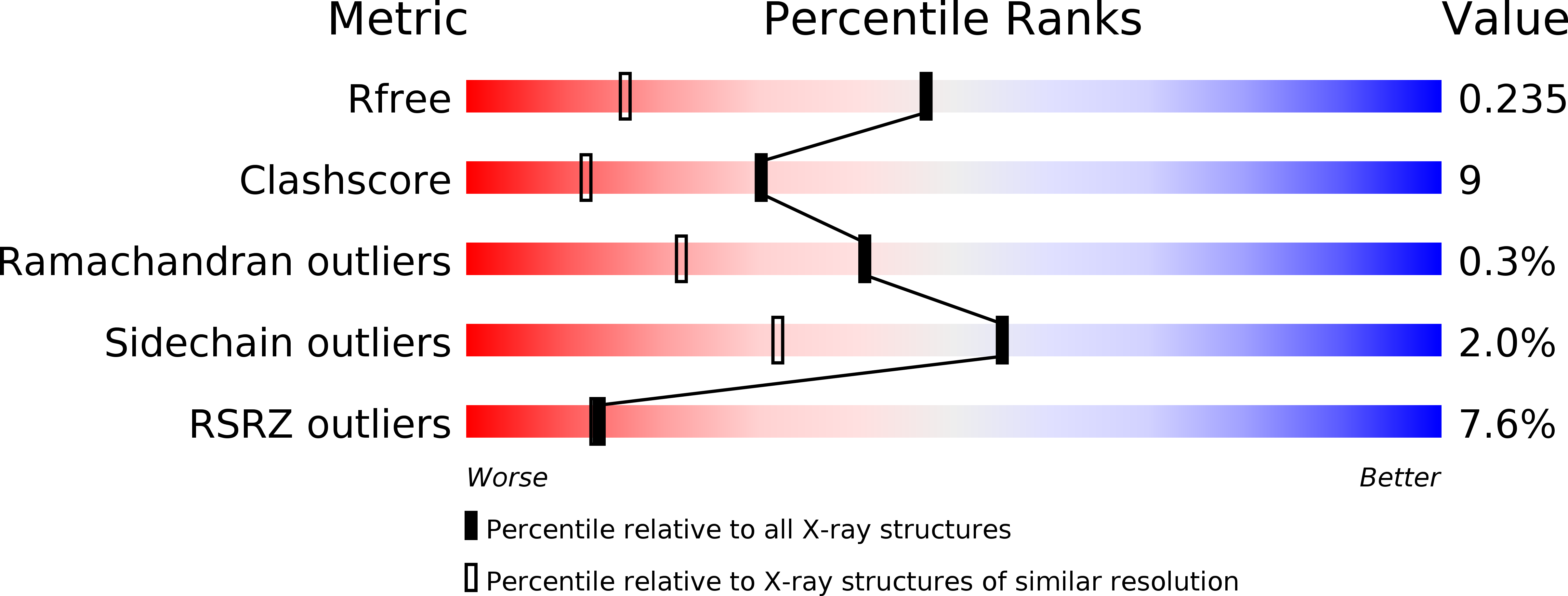
Resolution:
1.66 Å
R-Value Free:
0.24
R-Value Work:
0.20
R-Value Observed:
0.20
Space Group:
P 1 21 1
Macromolecular Entities
Structures with similar UniProt ID
Protein Blast
Polymer Type:polypeptide(L)
Molecule:Catabolite gene activator
Gene (Uniprot):crp
Mutagens:D138L
Chain IDs:A, B
Chain Length:260
Number of Molecules:2
Biological Source:Escherichia coli
Ligand Molecules
Primary Citation
The 1.6A resolution structure of activated D138L mutant of catabolite gene activator protein with two cAMP bound in each monomer
Int.J.Biol.Macromol. 48 459 465 (2011)
PMID: 21255606 DOI: 10.1016/j.ijbiomac.2011.01.009

Abstact

The X-ray crystal structure of the cAMP-liganded D138L mutant of Escherichia coli catabolite gene activator protein (CAP) was determined at a resolution of 1.66Å. This high resolution crystal structure reveals four cAMP binding sites in the homodimer. Two anti conformations of cAMPs (anti-cAMP) locate between the β-barrel and the C-helix of each subunit; two syn conformations of cAMPs (syn-cAMP) bind on the surface of the C-terminal domain. With two syn-cAMP molecules bound, the D138L CAP is highly symmetrical with both subunits assuming a "closed" conformation. These differences make the hinge region of the mutant more flexible. Protease susceptibility measurements indicate that D138L is more susceptible to proteases than that of wild type (WT) CAP. The results of protein dynamic experiments (H/D exchange measurements) indicate that the structure of D138L mutant is more dynamic than that of WT CAP, which may impact the recognition of specific DNA sequences.

Legend

Protein

Chemical

Disease

Primary Citation of related structures