Deposition Date
2009-10-20
Release Date
2010-02-02
Last Version Date
2023-09-06
Entry Detail
PDB ID:
3KBU
Keywords:
Title:
Crystal structure of the ankyrin binding domain of human erythroid beta spectrin (repeats 13-15) in complex with the spectrin binding domain of human erythroid ankyrin (ZU5-ANK), EMTS derivative
Biological Source:
Source Organism(s):
Homo sapiens (Taxon ID: 9606)
Expression System(s):
Method Details:
Experimental Method:
Resolution:
2.75 Å
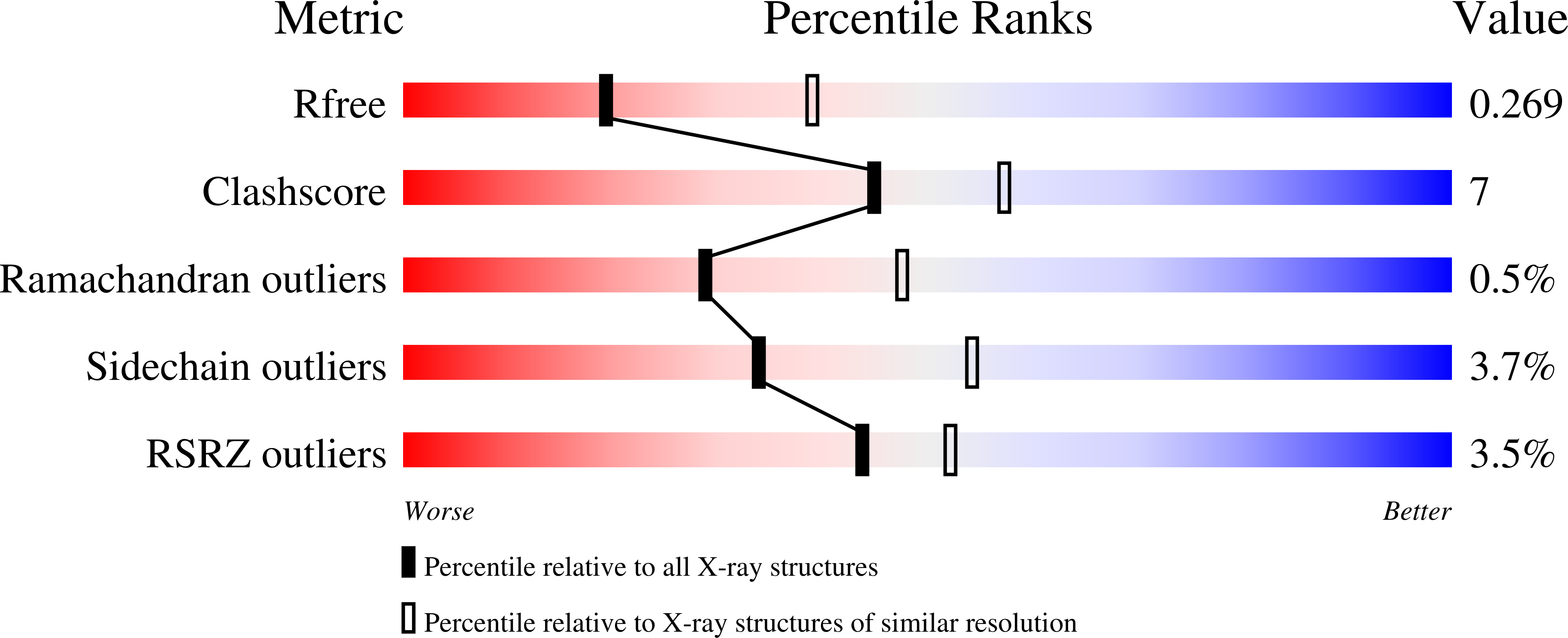
R-Value Free:
0.27
R-Value Work:
0.22
R-Value Observed:
0.22
Space Group:
P 21 21 21