Deposition Date
2009-10-20
Release Date
2009-11-10
Last Version Date
2023-11-15
Entry Detail
PDB ID:
3KB6
Keywords:
Title:
Crystal structure of D-Lactate dehydrogenase from aquifex aeolicus complexed with NAD and Lactic acid
Biological Source:
Source Organism(s):
Aquifex aeolicus (Taxon ID: 63363)
Expression System(s):
Method Details:
Experimental Method:
Resolution:
2.12 Å
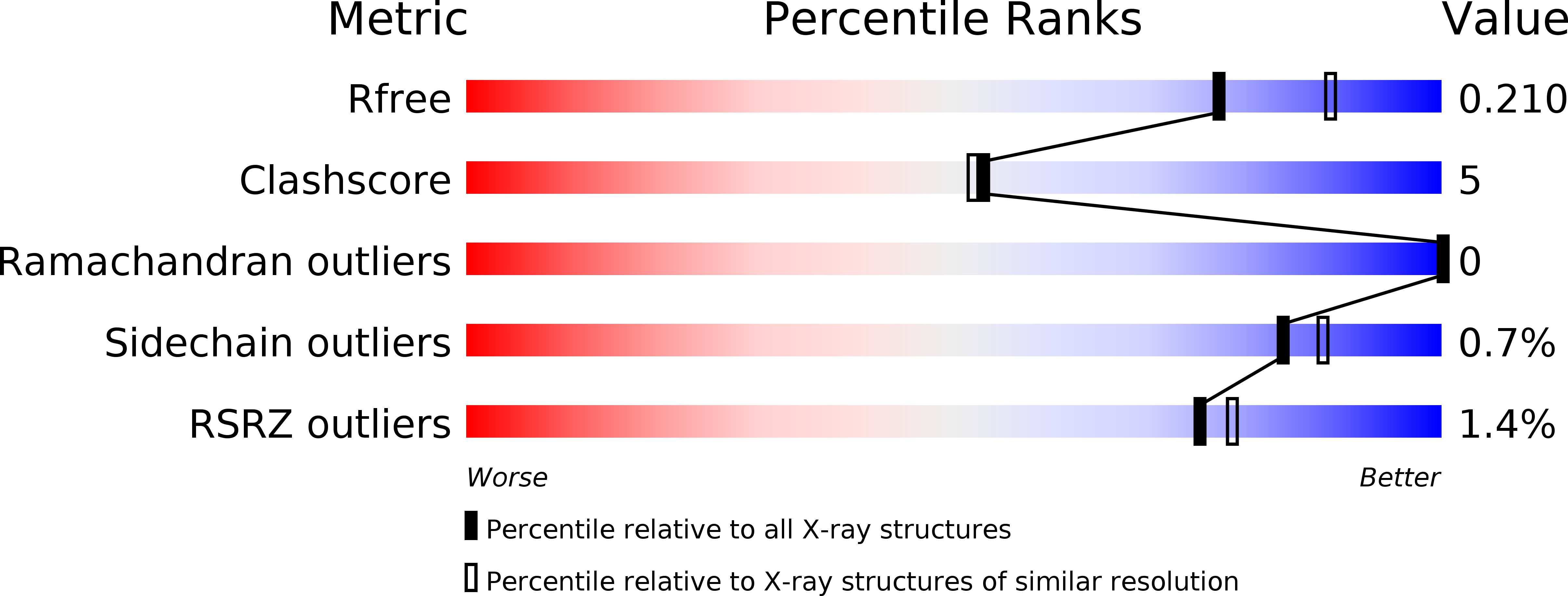
R-Value Free:
0.21
R-Value Work:
0.16
R-Value Observed:
0.16
Space Group:
P 21 21 21