3K8E image
Deposition Date 2009-10-14
Release Date 2009-11-10
Last Version Date 2023-11-01
Entry Detail
PDB ID:
3K8E
Keywords:
Title:
Crystal structure of E. coli lipopolysaccharide specific CMP-KDO synthetase
Biological Source:
Source Organism:
Method Details:
Experimental Method:
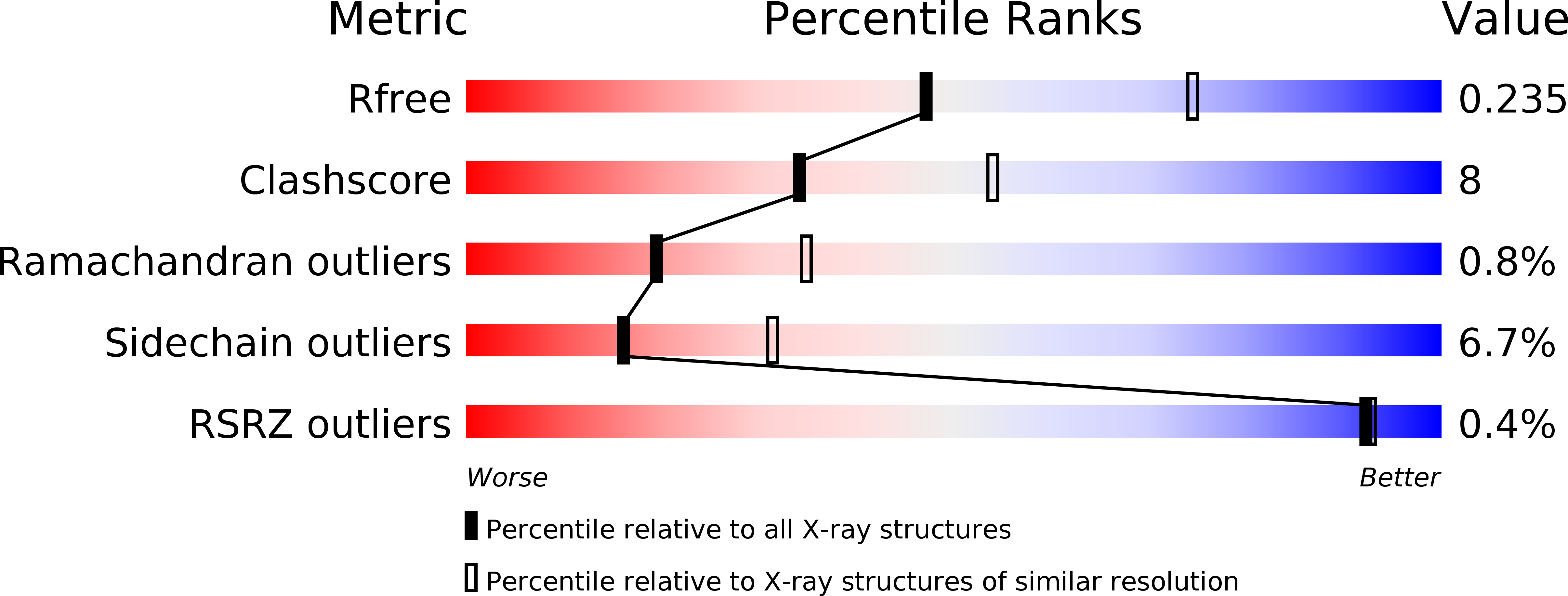
Resolution:
2.51 Å
R-Value Free:
0.23
R-Value Work:
0.19
R-Value Observed:
0.20
Space Group:
C 1 2 1
Macromolecular Entities
Structures with similar UniProt ID
Protein Blast
Polymer Type:polypeptide(L)
Molecule:3-deoxy-manno-octulosonate cytidylyltransferase
Gene (Uniprot):kdsB
Chain IDs:A (auth: C), B (auth: D), C (auth: A), D (auth: B)
Chain Length:264
Number of Molecules:4
Biological Source:Escherichia coli
Primary Citation
Structure-based mechanism of CMP-2-keto-3-deoxymanno-octulonic acid synthetase: convergent evolution of a sugar-activating enzyme with DNA/RNA polymerases
J.Biol.Chem. 284 35514 35523 (2009)
PMID: 19815542 DOI: 10.1074/jbc.M109.056630

Abstact

The enzyme CMP-Kdo synthetase (KdsB) catalyzes the addition of 2-keto-3-deoxymanno-octulonic acid (Kdo) to CTP to form CMP-Kdo, a key reaction in the biosynthesis of lipopolysaccharide. The reaction catalyzed by KdsB and the related CMP-acylneuraminate synthase is unique among the sugar-activating enzymes in that the respective sugars are directly coupled to a cytosine monophosphate. Using inhibition studies, in combination with isothermal calorimetry, we show the substrate analogue 2beta-deoxy-Kdo to be a potent competitive inhibitor. The ligand-free Escherichia coli KdsB and ternary complex KdsB-CTP-2beta-deoxy-Kdo crystal structures reveal that Kdo binding leads to active site closure and repositioning of the CTP phosphates and associated Mg(2+) ion (Mg-B). Both ligands occupy conformations compatible with an S(n)2-type attack on the alpha-phosphate by the Kdo 2-hydroxyl group. Based on strong similarity with DNA/RNA polymerases, both in terms of overall chemistry catalyzed as well as active site configuration, we postulate a second Mg(2+) ion (Mg-A) is bound by the catalytically competent KdsB-CTP-Kdo ternary complex. Modeling of this complex reveals the Mg-A coordinated to the conserved Asp(100) and Asp(235) in addition to the CTP alpha-phosphate and both the Kdo carboxylic and 2-hydroxyl groups. EPR measurements on the Mn(2+)-substituted ternary complex support this model. We propose the KdsB/CNS sugar-activating enzymes catalyze the formation of activated sugars, such as the abundant CMP-5-N-acetylneuraminic acid, by recruitment of two Mg(2+) to the active site. Although each metal ion assists in correct positioning of the substrates and activation of the alpha-phosphate, Mg-A is responsible for activation of the sugar-hydroxyl group.

Legend

Protein

Chemical

Disease

Primary Citation of related structures