3K7K image
Deposition Date 2009-10-13
Release Date 2010-01-26
Last Version Date 2023-11-01
Entry Detail
PDB ID:
3K7K
Keywords:
Title:
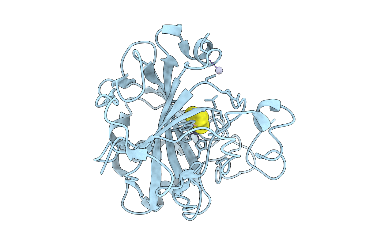
Crystal structure of the complex between Carbonic Anhydrase II and anions
Biological Source:
Source Organism:
Homo sapiens (Taxon ID: 9606)
Method Details:
Experimental Method:
Resolution:
1.90 Å
R-Value Free:
0.24
R-Value Work:
0.19
R-Value Observed:
0.19
Space Group:
P 1 21 1
Macromolecular Entities
Structures with similar UniProt ID
Protein Blast
Polymer Type:polypeptide(L)
Molecule:Carbonic anhydrase 2
Gene (Uniprot):CA2
Chain IDs:A
Chain Length:260
Number of Molecules:1
Biological Source:Homo sapiens
Primary Citation
Carbonic anhydrase inhibitors. X-ray crystal studies of the carbonic anhydrase II-trithiocarbonate adduct-An inhibitor mimicking the sulfonamide and urea binding to the enzyme
Bioorg.Med.Chem.Lett. 20 474 478 (2010)
PMID: 20005709 DOI: 10.1016/j.bmcl.2009.11.124

Abstact

Trithiocarbonate (CS32-) inhibits with low micromolar affinities several mammalian carbonic anhydrases, CAs, EC 4.2.1.1 [Innocenti et al., Bioorg. Med. Chem. Lett. 2009, 19, 1855]. Here we report the X-ray crystal structure of the hCA II-trithiocarbonate adduct. Trithiocarbonate is monodentately bound to the Zn(II) ion and makes several hydrogen bonds with Thr199 and two water molecules from the enzyme active site. Its binding is different from that of ureate, another small inhibitor isosteric with trithiocarbonate but somehow mimicks the binding of the SO(2)NH moiety present in the sulfonamide inhibitors and is similar to that of bicarbonate. Compounds incorporating this new zinc-binding group, CS2-, may thus lead to new classes of potent inhibitors.

Legend

Protein

Chemical

Disease

Primary Citation of related structures