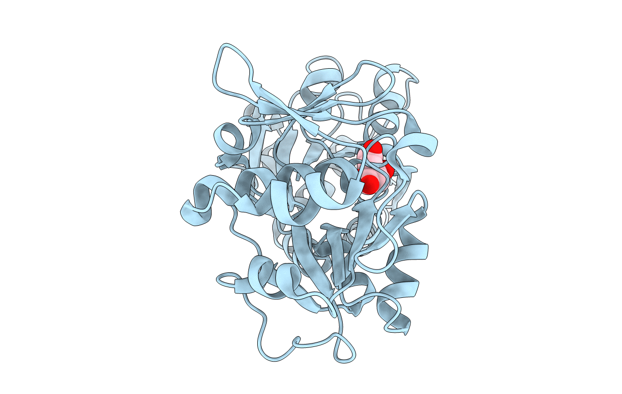
3K6V image
Entry Detail
PDB ID:
3K6V
Title:
M. acetivorans Molybdate-Binding Protein (ModA) in Citrate-Bound Open Form
Biological Source:
Host Organism:
PDB Version:
Deposition Date:
2009-10-09
Release Date:
2010-01-12
Method Details:
Experimental Method:
Resolution:
1.69 Å
R-Value Free:
0.21
R-Value Work:
0.18
R-Value Observed:
0.19
Space Group:
P 61
Macromolecular Entities
Polymer Type:polypeptide(L)
Description:Solute-binding protein MA_0280
Chain IDs:A
Chain Length:354
Number of Molecules:1
Biological Source:Methanosarcina acetivorans
Ligand Molecules
Primary Citation
Apo and ligand-bound structures of ModA from the archaeon Methanosarcina acetivorans
Acta Crystallogr.,Sect.F 66 242 250 (2010)
PMID: 20208152 DOI: 10.1107/S1744309109055158

Abstact

The trace-element oxyanion molybdate, which is required for the growth of many bacterial and archaeal species, is transported into the cell by an ATP-binding cassette (ABC) transporter superfamily uptake system called ModABC. ModABC consists of the ModA periplasmic solute-binding protein, the integral membrane-transport protein ModB and the ATP-binding and hydrolysis cassette protein ModC. In this study, X-ray crystal structures of ModA from the archaeon Methanosarcina acetivorans (MaModA) have been determined in the apoprotein conformation at 1.95 and 1.69 A resolution and in the molybdate-bound conformation at 2.25 and 2.45 A resolution. The overall domain structure of MaModA is similar to other ModA proteins in that it has a bilobal structure in which two mixed alpha/beta domains are linked by a hinge region. The apo MaModA is the first unliganded archaeal ModA structure to be determined: it exhibits a deep cleft between the two domains and confirms that upon binding ligand one domain is rotated towards the other by a hinge-bending motion, which is consistent with the 'Venus flytrap' model seen for bacterial-type periplasmic binding proteins. In contrast to the bacterial ModA structures, which have tetrahedral coordination of their metal substrates, molybdate-bound MaModA employs octahedral coordination of its substrate like other archaeal ModA proteins.

Legend

Protein

Chemical

Disease

Primary Citation of related structures