3K6T image
Deposition Date 2009-10-09
Release Date 2010-03-09
Last Version Date 2024-02-21
Entry Detail
PDB ID:
3K6T
Keywords:
Title:
Crystal structure of the GLD-1 homodimerization domain from Caenorhabditis elegans at 2.04 A resolution
Biological Source:
Source Organism:
Host Organism:
Method Details:
Experimental Method:
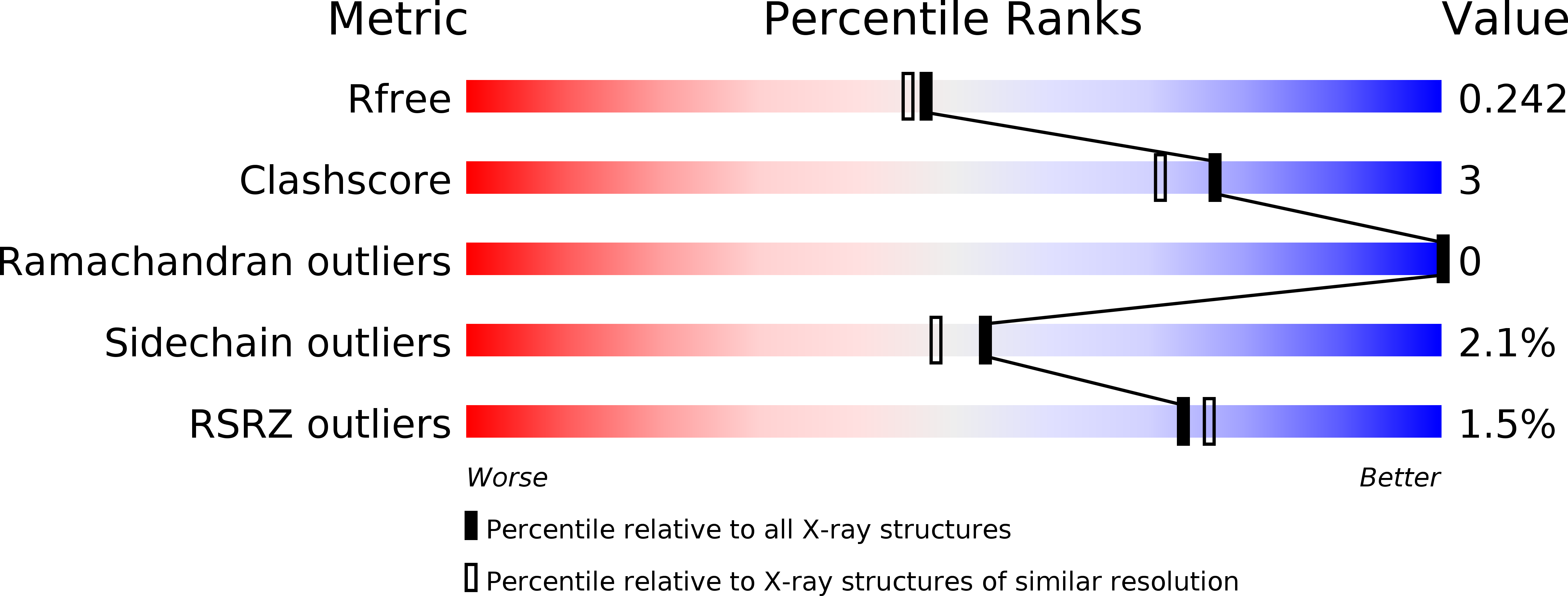
Resolution:
2.04 Å
R-Value Free:
0.24
R-Value Work:
0.19
R-Value Observed:
0.20
Space Group:
P 21 21 21
Macromolecular Entities
Polymer Type:polypeptide(L)
Molecule:Female germline-specific tumor suppressor gld-1
Gene (Uniprot):gld-1
Chain IDs:A, B, C, D
Chain Length:60
Number of Molecules:4
Biological Source:Caenorhabditis elegans
Primary Citation
Structure of the GLD-1 homodimerization domain: insights into STAR protein-mediated translational regulation.
Structure 18 377 389 (2010)
PMID: 20223220 DOI: 10.1016/j.str.2009.12.016

Abstact

Posttranscriptional regulation of gene expression is an important mechanism for modulating protein levels in eukaryotes, especially in developmental pathways. The highly conserved homodimeric STAR/GSG proteins play a key role in regulating translation by binding bipartite consensus sequences in the untranslated regions of target mRNAs, but the exact mechanism remains unknown. Structures of STAR protein RNA binding subdomains have been determined, but structural information is lacking for the homodimerization subdomain. Here, we present the structure of the C. elegans GLD-1 homodimerization domain dimer, determined by a combination of X-ray crystallography and NMR spectroscopy, revealing a helix-turn-helix monomeric fold with the two protomers stacked perpendicularly. Structure-based mutagenesis demonstrates that the dimer interface is not easily disrupted, but the structural integrity of the monomer is crucial for GLD-1 dimerization. Finally, an improved model for STAR-mediated translational regulation of mRNA, based on the GLD-1 homodimerization domain structure, is presented.

Legend

Protein

Chemical

Disease

Primary Citation of related structures