Deposition Date
2009-10-09
Release Date
2009-11-17
Last Version Date
2023-09-06
Entry Detail
PDB ID:
3K6N
Keywords:
Title:
Crystal structure of the S225E mutant Kir3.1 cytoplasmic pore domain
Biological Source:
Source Organism(s):
Mus musculus (Taxon ID: 10090)
Expression System(s):
Method Details:
Experimental Method:
Resolution:
2.00 Å
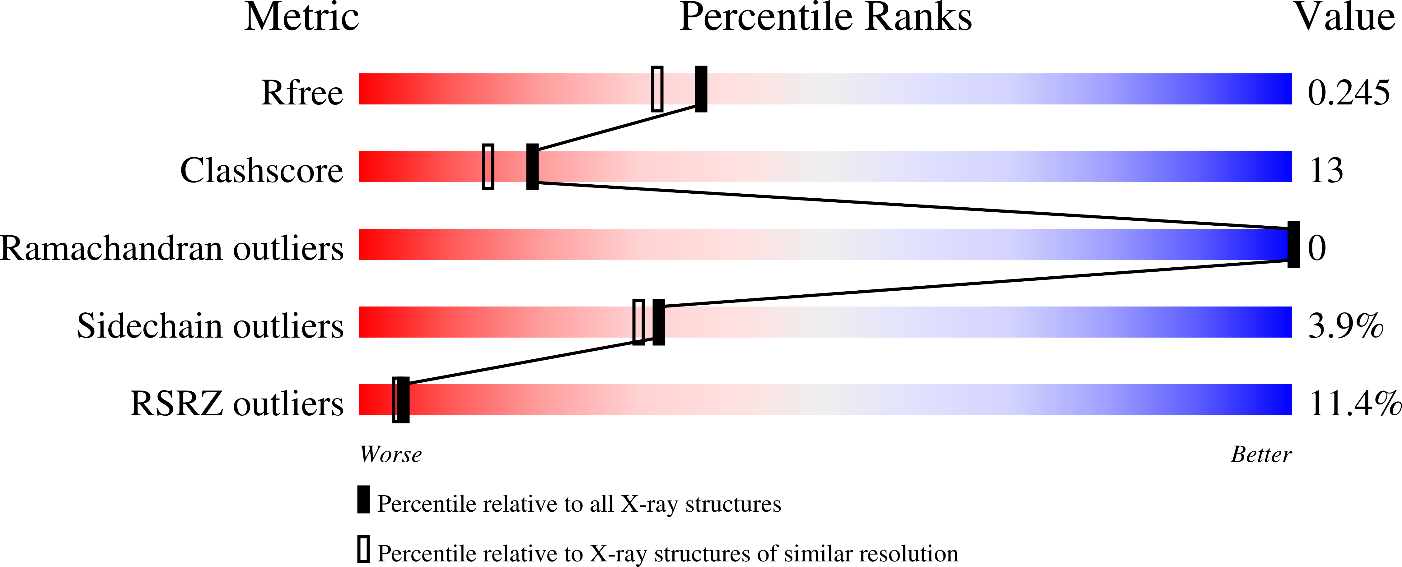
R-Value Free:
0.26
R-Value Work:
0.25
Space Group:
P 4 21 2