3K6B image
Deposition Date 2009-10-08
Release Date 2009-11-10
Last Version Date 2023-09-06
Entry Detail
PDB ID:
3K6B
Keywords:
Title:
X-ray crystal structure of the E2 domain of APL-1 from C. elegans, in complex with sucrose octasulfate (SOS)
Biological Source:
Source Organism:
Host Organism:
Method Details:
Experimental Method:
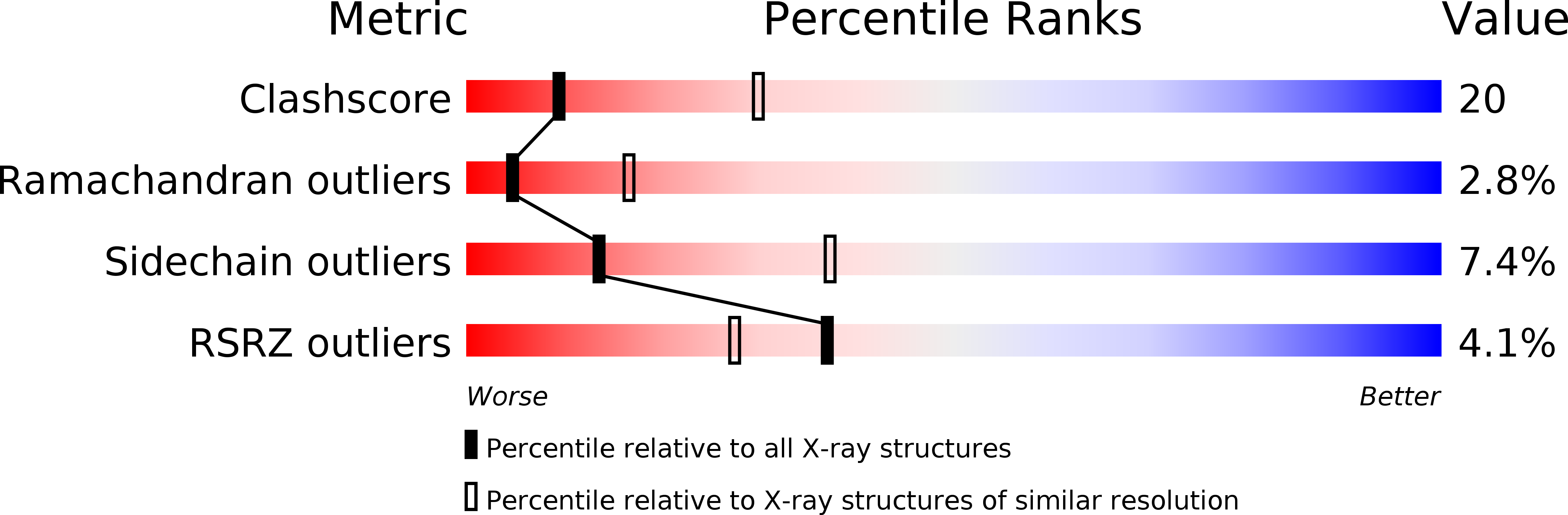
Resolution:
2.80 Å
R-Value Free:
0.30
R-Value Work:
0.26
R-Value Observed:
0.26
Space Group:
P 32 2 1
Macromolecular Entities
Polymer Type:polypeptide(L)
Molecule:Beta-amyloid-like protein
Gene (Uniprot):apl-1
Chain IDs:A
Chain Length:239
Number of Molecules:1
Biological Source:Caenorhabditis elegans
Ligand Molecules
Primary Citation
Structural characterization of the E2 domain of APL-1, a Caenorhabditis elegans homolog of human amyloid precursor protein, and its heparin binding site
J.Biol.Chem. 285 2165 2173 (2010)
PMID: 19906646 DOI: 10.1074/jbc.M109.018432

Abstact

The amyloid beta-peptide deposit found in the brain tissue of patients with Alzheimer disease is derived from a large heparin-binding protein precursor APP. The biological function of APP and its homologs is not precisely known. Here we report the x-ray structure of the E2 domain of APL-1, an APP homolog in Caenorhabditis elegans, and compare it to the human APP structure. We also describe the structure of APL-1 E2 in complex with sucrose octasulfate, a highly negatively charged disaccharide, which reveals an unexpected binding pocket between the two halves of E2. Based on the crystal structure, we are able to map, using site-directed mutagenesis, a surface groove on E2 to which heparin may bind. Our biochemical data also indicate that the affinity of E2 for heparin is influenced by pH: at pH 5, the binding appears to be much stronger than that at neutral pH. This property is likely caused by histidine residues in the vicinity of the mapped heparin binding site and could be important for the proposed adhesive function of APL-1.

Legend

Protein

Chemical

Disease

Primary Citation of related structures