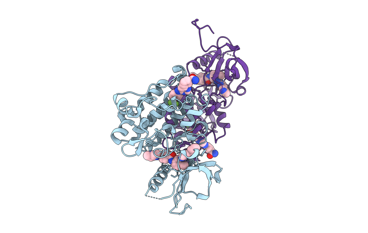
3K5V image
Entry Detail
PDB ID:
3K5V
Keywords:
Title:
Structure of Abl kinase in complex with imatinib and GNF-2
Biological Source:
Source Organism:
Host Organism:
PDB Version:
Deposition Date:
2009-10-08
Release Date:
2010-01-19
Method Details:
Experimental Method:
Resolution:
1.74 Å
R-Value Free:
0.23
R-Value Work:
0.19
R-Value Observed:
0.20
Space Group:
P 1
Macromolecular Entities
Polymer Type:polypeptide(L)
Description:Tyrosine-protein kinase ABL1
Chain IDs:A, B
Chain Length:293
Number of Molecules:2
Biological Source:Mus musculus
Primary Citation

Abstact

In an effort to find new pharmacological modalities to overcome resistance to ATP-binding-site inhibitors of Bcr-Abl, we recently reported the discovery of GNF-2, a selective allosteric Bcr-Abl inhibitor. Here, using solution NMR, X-ray crystallography, mutagenesis and hydrogen exchange mass spectrometry, we show that GNF-2 binds to the myristate-binding site of Abl, leading to changes in the structural dynamics of the ATP-binding site. GNF-5, an analogue of GNF-2 with improved pharmacokinetic properties, when used in combination with the ATP-competitive inhibitors imatinib or nilotinib, suppressed the emergence of resistance mutations in vitro, displayed additive inhibitory activity in biochemical and cellular assays against T315I mutant human Bcr-Abl and displayed in vivo efficacy against this recalcitrant mutant in a murine bone-marrow transplantation model. These results show that therapeutically relevant inhibition of Bcr-Abl activity can be achieved with inhibitors that bind to the myristate-binding site and that combining allosteric and ATP-competitive inhibitors can overcome resistance to either agent alone.

Legend

Protein

Chemical

Disease

Primary Citation of related structures