3K5J image
Entry Detail
PDB ID:
3K5J
Keywords:
Title:
Crystal structure of Putative SUFU (suppressor of fused protein) homolog (YP_208451.1) from Neisseria gonorrhoeae FA 1090 at 1.40 A resolution
Biological Source:
Source Organism:
Host Organism:
PDB Version:
Deposition Date:
2009-10-07
Release Date:
2010-01-26
Method Details:
Experimental Method:
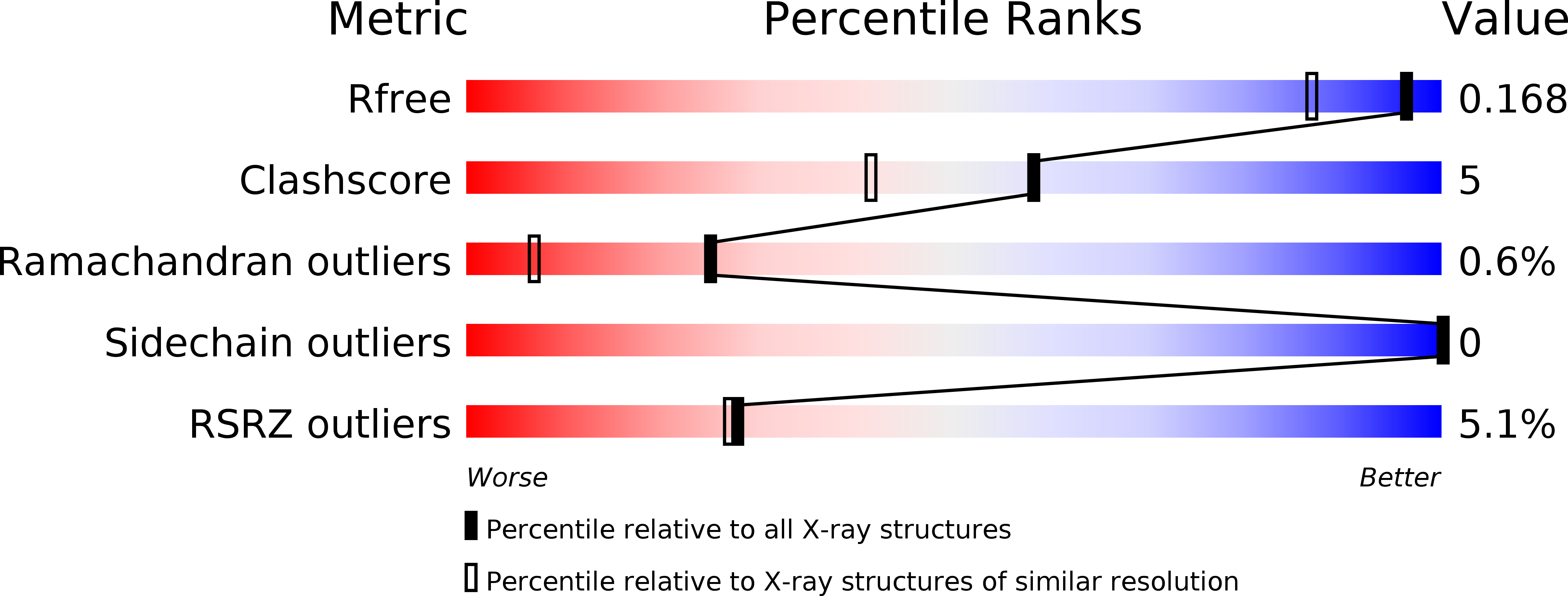
Resolution:
1.40 Å
R-Value Free:
0.16
R-Value Work:
0.13
R-Value Observed:
0.13
Space Group:
P 32 2 1
Macromolecular Entities
Polymer Type:polypeptide(L)
Description:Suppressor of fused family protein
Chain IDs:A
Chain Length:183
Number of Molecules:1
Biological Source:Neisseria gonorrhoeae
Modified Residue
Compound ID Chain ID Parent Comp ID Details 2D Image
MSE A MET modified residue
Primary Citation

Abstact

Sufu (Suppressor of Fused), a two-domain protein, plays a critical role in regulating Hedgehog signaling and is conserved from flies to humans. A few bacterial Sufu-like proteins have previously been identified based on sequence similarity to the N-terminal domain of eukaryotic Sufu proteins, but none have been structurally or biochemically characterized and their function in bacteria is unknown. We have determined the crystal structure of a more distantly related Sufu-like homolog, NGO1391 from Neisseria gonorrhoeae, at 1.4 Å resolution, which provides the first biophysical characterization of a bacterial Sufu-like protein. The structure revealed a striking similarity to the N-terminal domain of human Sufu (r.m.s.d. of 2.6 Å over 93% of the NGO1391 protein), despite an extremely low sequence identity of ∼15%. Subsequent sequence analysis revealed that NGO1391 defines a new subset of smaller, Sufu-like proteins that are present in ∼200 bacterial species and has resulted in expansion of the SUFU (PF05076) family in Pfam.

Legend

Protein

Chemical

Disease

Primary Citation of related structures